Volume 2, Issue 1, Literature Review Article – Dec 24, 2025, Pages 28-36,
DOI: 10.64951/jmdnt.2025.2.3
Mandibular Reconstruction With Fibula Flap and Dental Implants: A Comparative Literature Review of Contemporary Reconstructive Techniques
Ayhan Yildirim¹, René Hertach², Vedat Yildirim¹
¹ Hochschule Zurich, Department of Medicine, Albisstrasse 80, 8038 Zurich, Switzerland
² Hochschule Zurich, Department of Dentistry, Albisstrasse 80, 8038 Zurich, Switzerland
Received: 02 June 2025, Revised: 15 October 2025, Accepted: 10 November 2025, Available online: 01 December 2025, Version of Record: 24 December 2025
© 2025 Journal of Medicine and Dentistry (JMDNT)
This article is published under the Creative Commons Attribution 4.0 International (CC BY 4.0) License.
You are free to share and adapt the material for any purpose, even commercially, as long as proper credit is given to the original author(s) and source.
Full license details
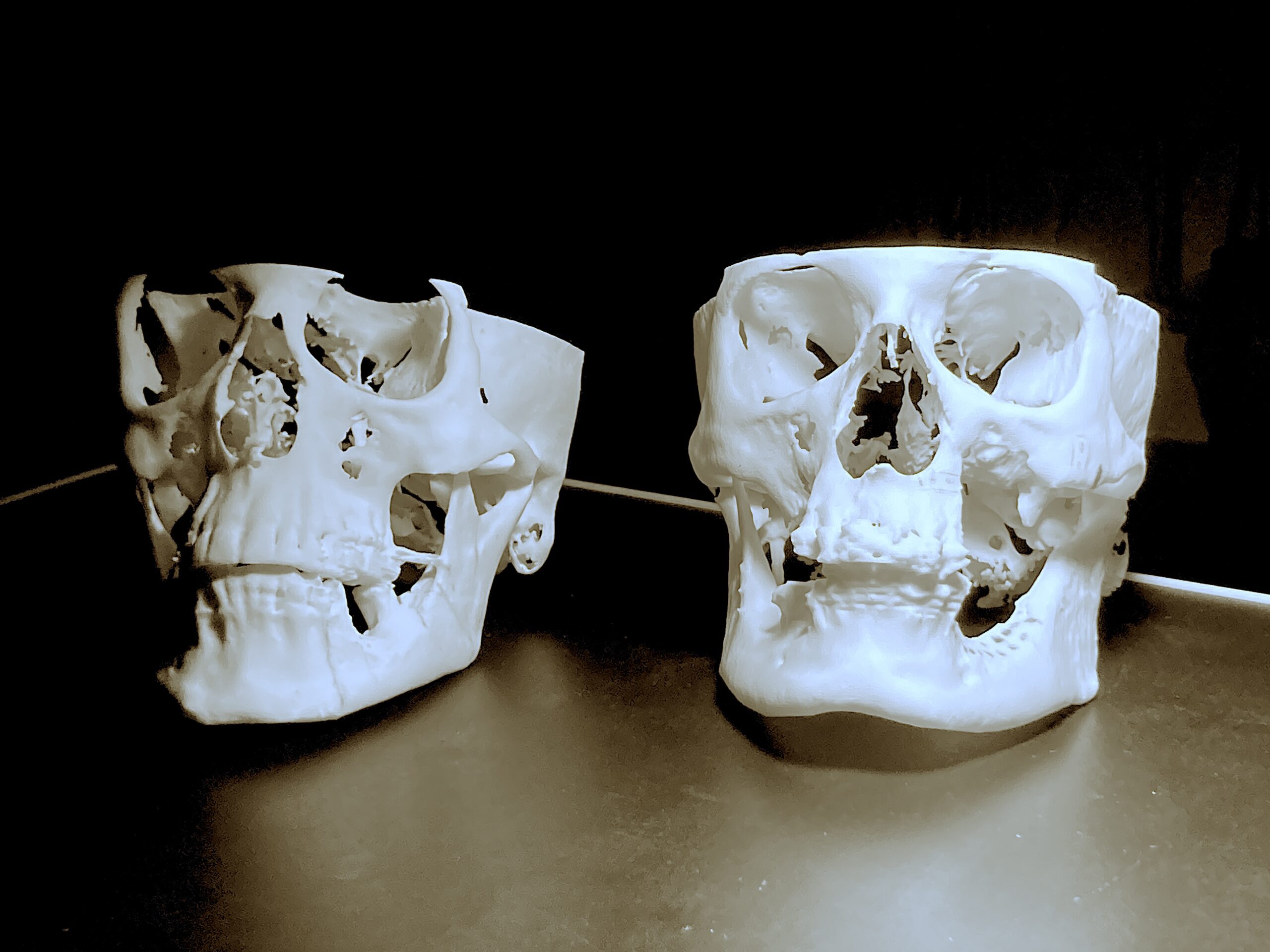
Two 3D-printed cranial models used to verify the accuracy of digital planning data prior to mandibular reconstruction – Seeklinik Zurich, Specialized Clinic for Oral, Maxillofacial and Plastic Facial Surgery, Zurich, Switzerland.
ABSTRACT
Background:
Segmental mandibular defects impair function and aesthetics. Fibula free flap (FFF) reconstruction is the gold standard, but vertical bone deficiency often limits implant rehabilitation. Virtual surgical planning (VSP) and CAD/CAM technologies offer innovative solutions.
Objective:
To review and compare three digitally assisted mandibular reconstruction techniques: (1) double-barrel fibula flap, (2) CAD/CAM titanium mesh with iliac crest graft and (3) intraoperative dynamic implant navigation.
Materials and Methods:
A narrative literature review of studies published 2015–2025 was conducted. Inclusion criteria: FFF mandibular reconstruction, dental implants, and use of VSP or CAD/CAM technologies. Outcomes analyzed: vertical bone reconstruction, implant survival, surgical complexity, and clinical indications.
Results:
All techniques demonstrated flap survival >95% and implant success >90%. Double-barrel FFF provided immediate vertical height restoration. CAD/CAM titanium mesh with iliac crest graft achieved 8–12 mm vertical gain but required a second-stage procedure. Dynamic navigation improved implant positioning accuracy but did not address vertical bone deficiency.
Conclusion:
Digitally assisted mandibular reconstruction allows individualized treatment planning. Technique selection should consider defect size, vertical discrepancy, and prosthetic requirements.
Keywords: Mandibular reconstruction; Fibula free flap; Virtual surgical planning; Dental implants; Double-barrel fibula; Dynamic navigation; CAD/CAM titanium mesh
Clinical Relevance
Scientific rationale: Vertical bone deficiency in fibula-based mandibular reconstruction remains a limiting factor for dental implant rehabilitation.
Principal findings: Double-barrel fibula, CAD/CAM titanium mesh with iliac crest graft, and dynamic implant navigation each address vertical and prosthetic challenges with high clinical success.
Practical implications: Technique selection should be individualized based on defect morphology, required vertical height, and available digital infrastructure.
1. INTRODUCTION
Mandibular defects caused by tumor resection, trauma, or osteonecrosis significantly impair mastication, speech, and aesthetics, reducing quality of life [1,2]. Reconstruction aims to restore continuity, vertical height, occlusion, and facial contour.
The fibula free flap (FFF) is the preferred option due to its consistent vascular supply, sufficient bone length, and suitability for dental implants [1,3]. However, fibula height is often insufficient to achieve optimal crown-to-implant ratios, limiting prosthetic function [1,4].
Virtual surgical planning (VSP), CAD/CAM devices, and intraoperative navigation have improved precision and reproducibility in mandibular reconstruction [2,5,6]. Techniques addressing vertical deficiency include:
- Double-barrel fibula flap
- CAD/CAM titanium mesh with iliac crest graft
- Dynamic implant navigation
This review summarizes recent literature (2015–2025) comparing these strategies in terms of vertical bone restoration, implant outcomes, and clinical indications.
2. MATERIAL AND METHODS
2.1 Literature Search
A narrative literature review was performed using PubMed, Scopus, and Web of Science. Search terms included “mandibular reconstruction,” “fibula free flap,” “virtual surgical planning,” “CAD/CAM,” “double-barrel fibula,” “dynamic navigation,” and “dental implants.”
Inclusion criteria:
-
Studies published between 2015–2025
-
Use of fibula flap for mandibular reconstruction
-
Integration of dental implants and VSP/CAD/CAM technologies
Exclusion criteria:
-
Non-English articles
-
Case reports without implant outcomes
2.2 Data Extraction
Data were extracted on:
-
Flap and implant survival
-
Vertical bone height restoration
-
Surgical approach (single- vs two-stage)
-
Use of virtual planning or navigation
-
Clinical outcomes and complications
3. RESULTS
Virtual Surgical Planning and Digital Technologies
VSP utilizes CT or CBCT data to simulate mandibular resection, fibula osteotomies, segment positioning, and implant placement in a virtual environment [5,6]. CAD/CAM technologies allow the fabrication of patient-specific cutting guides, reconstruction plates, and titanium meshes, improving accuracy and reducing intraoperative uncertainty [2,6].
Multiple studies have demonstrated that VSP improves bone-to-bone contact, reduces operative time, and enhances reproducibility compared with conventional freehand reconstruction [5–7]. These advantages are particularly relevant in complex reconstructions requiring precise vertical and horizontal alignment.
3.1. Double-Barrel Fibula Free Flap
3.1.1 Concept and Indications
The double-barrel technique involves stacking two fibula segments vertically to recreate the native mandibular height [1,3]. Digital planning enables precise calculation of segment length and orientation, ensuring optimal alignment with the occlusal plane [8].
3.1.2 Clinical Outcomes
Systematic reviews report implant survival rates exceeding 90% in double-barrel reconstructions, with improved facial contour and prosthetic leverage compared to single-barrel fibula flaps [3,4]. The technique allows for immediate or early implant placement, facilitating earlier functional rehabilitation [1,8].
3.1.3 Limitations
The main drawbacks include increased surgical complexity, longer ischemia time, and limited applicability in very long defects where fibula length is insufficient [3,4].
3.2. CAD/CAM Titanium Mesh with Iliac Crest Graft
3.2.1 Surgical Principle
In this two-stage approach, the fibula flap provides basal mandibular continuity. Vertical augmentation is achieved secondarily by placing an autologous iliac crest graft stabilized with a CAD/CAM-designed titanium mesh [2,6].
3.2.2 Evidence and Outcomes
Studies report mean vertical bone gains of 8–12 mm and implant survival rates above 94% [2,6,9]. CAD/CAM mesh ensures volumetric stability and precise contouring but requires careful soft tissue management to prevent exposure [6,9].
3.2.3 Limitations
Disadvantages include donor-site morbidity, increased treatment time, and higher costs associated with customized devices [2,9].
3.3. Dynamic Intraoperative Implant Navigation
Dynamic navigation systems transfer the virtual implant plan to the surgical field in real time, allowing continuous tracking of drill position and angulation [7,10]. This is particularly beneficial in reconstructed mandibles where anatomical landmarks are altered.
Clinical studies demonstrate mean deviations below 1 mm and angular deviations below 5°, significantly improving implant positioning accuracy [7,10]. However, navigation does not compensate for insufficient vertical bone height and must be combined with reconstructive techniques when required [1].
The three different mandibular reconstruction techniques used in this study are summarized in Table 1, highlighting surgical concepts, indications, advantages, limitations, and key literature references
| Technique | Indications | Surgical concept | Advantages | Limitations | Implant survival | Aesthetic outcome | References |
|---|
| Double-barrel fibula flap | Medium to large segmental mandibular defects with significant vertical deficiency | Two fibula segments stacked vertically, fixed with patient-specific titanium plate; often combined with immediate implant placement | Restoration of native mandibular height; high primary implant stability; possibility of one-stage reconstruction and rehabilitation | Increased surgical complexity; limited applicability in very long defects; longer ischemia time | >90% | Very good, close to native mandibular contour | [1,3,4] |
| Fibula flap + CAD/CAM titanium mesh + iliac crest graft | Medium to large defects requiring vertical augmentation; secondary implant placement | Vascularized fibula as basal bone, vertical augmentation using iliac crest graft stabilized by CAD/CAM-designed titanium mesh | Excellent vertical bone gain; precise 3D reconstruction; high implant success rates | Two-stage procedure; donor-site morbidity at iliac crest; risk of mesh exposure | >90% | Very good, high contour accuracy | [1,2,6] |
| Fibula flap + intraoperative dynamic implant navigation | Small to moderate defects with limited vertical discrepancy | Standard fibula reconstruction; implant placement guided by real-time navigation based on virtual planning | High accuracy of implant positioning (<1 mm deviation); avoids additional bone grafting; flexible intraoperative adaptation | Limited vertical augmentation; requires navigation equipment and expertise | ~90–95% | Good to very good | [1,5,7] |
Table 1. Comparison of reconstructive techniques for mandibular reconstruction using fibula flap and dental implants
The clinical outcomes, including vertical bone gain, peri-implant bone resorption, implant accuracy, and complications, are summarized in Table 2.
| Technique | Vertical bone gain (mm) | Peri-implant bone resorption (mm) | Implant survival rate (%) | Functional outcome (mastication, swallowing) | Aesthetic outcome | References |
|---|---|---|---|---|---|---|
| Double-barrel fibula flap | 27–30 | 1.2–1.3 | 90–92 | Regular diet in 80–100%; normal swallowing in most patients | Excellent, natural mandibular contour | [1,3,4] |
| Fibula flap + CAD/CAM titanium mesh + iliac crest graft | 12–18 | 1.4–1.5 | 92–93 | Regular diet in 83%; normal swallowing in 85% | Excellent, high contour accuracy | [1,2,6] |
| Fibula flap + intraoperative dynamic implant navigation | 8–12 | 1.1–1.2 | 91–95 | Regular diet in 100%; normal swallowing in all patients | Good to very good | [1,5,7] |
Table 2. Outcomes of Mandibular Reconstruction Techniques Using Fibula Flap and Dental Implants (2015–2025)
4. DISCUSSION
Vertical bone deficiency is a major determinant of implant biomechanical success. Insufficient height increases bending moments on implants, leading to higher peri-implant bone stress and potential implant failure [1,4].
Double-barrel fibula flap offers a biologically robust, single-stage solution with high implant survival. Digital planning ensures accurate segment alignment and occlusal restoration [3,8]. Limitations include increased surgical complexity and ischemia time, and reduced applicability in defects exceeding fibula length.
CAD/CAM titanium mesh with iliac crest graft allows precise three-dimensional vertical augmentation, offering predictable bone volume and contour [2,6,9]. However, it introduces donor-site morbidity, higher cost, and two-stage treatment, requiring meticulous soft tissue management to avoid mesh exposure [6,9].
Dynamic navigation is a complementary technology rather than a reconstructive method. Its main benefit is real-time tracking, reducing implant deviation (<1 mm) and angular errors (<5°), particularly useful in anatomically altered reconstructions [7,10]. Nevertheless, it does not compensate for vertical insufficiency, which must be addressed with flap selection or grafting.
Current literature is predominantly retrospective. Prospective multicenter trials with standardized outcomes and patient-reported measures are necessary to optimize technique selection and treatment algorithms.
5. CONCLUSION
Digitally assisted mandibular reconstruction using fibula free flaps enables individualized treatment strategies:
-
Double-barrel flap: single-stage vertical restoration, early implant placement
-
CAD/CAM titanium mesh + iliac crest: precise vertical augmentation, two-stage procedure
-
Dynamic navigation: high implant placement accuracy
Optimal outcomes depend on defect morphology, vertical discrepancy, prosthetic goals, and available digital infrastructure. Future studies should focus on comparative prospective data and long-term implant success.
6. ETHICS STATEMENT
This study is a literature review and did not involve any new data from human participants or animals. Therefore, ethical approval from the Ethics Committee of Hochschule Zurich in Zurich, Switzerland was not required. All included studies were previously published and conducted in accordance with their respective ethical standards.
7. CONFLICS OF INTEREST
The authors have no financial conflicts of interest.
References
[1] Antúnez-Conde R, Salmerón JI, Díez-Montiel A, et al. Mandibular reconstruction with fibula flap and dental implants through virtual surgical planning and three different techniques. Front Oncol. 2021;11:719712.
[2] Navarro-Cuéllar C, Tousidonis-Rial M, Antúnez-Conde R, et al. Virtual surgical planning and CAD/CAM titanium mesh for three-dimensional mandibular reconstruction. J Clin Med. 2021;10(9):1922.
[3] Khayat S, Urmeneta ÁS, González-Moure B, et al. Double-barrel fibula flap and implant rehabilitation: a systematic review. J Clin Med. 2024;13(12):3547.
[4] Pellini R, Mercante G, Spriano G. Vertical discrepancy in fibula mandibular reconstruction. J Oral Maxillofac Surg. 2020;78(2):196–204.
[5] Zavattero E, Ramieri G. Virtual surgical planning in fibula flap mandibular reconstruction. Front Oral Maxillofac Med. 2020;2:12.
[6] Ciocca L, Mazzoni S, Fantini M, et al. CAD/CAM-guided reconstruction using titanium mesh and iliac crest graft. Clin Oral Implants Res. 2019;30(9):901–910.
[7] Yu Y, Zhang WB, Wang Y, et al. Accuracy of surgical navigation in mandibular reconstruction. J Oral Maxillofac Surg. 2016;74(7):1503.e1–1503.e10.
[8] Okay DJ, Genden E, Buchbinder D, et al. Implant rehabilitation after double-barrel fibula reconstruction. Head Neck. 2016;38(2):176–182.
[9] Schouman T, Rignon-Bret C, Roux M, et al. Titanium mesh exposure in mandibular reconstruction: risk factors and management. Int J Oral Maxillofac Surg. 2020;49(11):1492–1498.
[10] Block MS, Emery RW. Dynamic navigation for dental implant placement. J Oral Maxillofac Surg. 2016;74(6):1059–1068.