Volume 3, Issue 2, Prospective Comparative Validation Study – Apr 24, 2026, Pages 85-95
DOI: 10.64951/jmdnt.2026.2.22
Three-Dimensional Accuracy of AI-Assisted Virtual Surgical Planning in Bimaxillary Orthognathic Surgery
Ayhan Yildirim¹, René Hertach², Vedat Yildirim²
¹ Hochschule Zurich, Department of Medicine, Albisstrasse 80, 8038 Zurich, Switzerland
² Hochschule Zurich, Department of Dentistry, Albisstrasse 80, 8038 Zurich, Switzerland
Received: 21 January 2026, Revised: 05 March 2026, Accepted: 26 March, 2026, Available online: 24 April 2026, Version of Record: 24 April 2026
© 2026 Journal of Medicine and Dentistry (JMDNT)
This article is published under the Creative Commons Attribution 4.0 International (CC BY 4.0) License.
You are free to share and adapt the material for any purpose, even commercially, as long as proper credit is given to the original author(s) and source.
Full license details
ABSTRACT
Background
Artificial intelligence (AI), virtual surgical planning, and additive manufacturing increasingly influence modern orthognathic surgery workflows. Previous investigations demonstrated the feasibility and translational integration of AI-assisted workflows in maxillofacial trauma surgery and recently also in bimaxillary orthognathic surgery [1–13]. However, prospective validation data regarding the three-dimensional surgical transfer accuracy of AI-assisted planning workflows remain limited.
Objective
The aim of this study was to evaluate the three-dimensional surgical accuracy of AI-assisted virtual surgical planning and patient-specific 3D-printed splint transfer in bimaxillary orthognathic surgery compared with conventional planning workflows.
Methods
A prospective comparative validation study was conducted between January 2020 and December 2025 at Seeklinik Zurich, Switzerland. Consecutive patients undergoing bimaxillary orthognathic surgery were prospectively evaluated using postoperative three-dimensional superimposition analysis. Planned-versus-achieved skeletal movement accuracy, rotational and translational deviations, splint transfer precision, postoperative symmetry, and occlusal stability were assessed longitudinally.
Results
A total of 124 patients were included in the final analysis, including 66 patients treated using AI-assisted workflows and 58 patients treated using conventional planning methods. AI-assisted workflows demonstrated significantly reduced three-dimensional deviation values and improved skeletal transfer precision compared with conventional planning. Maxillary and mandibular repositioning accuracy showed lower translational and rotational discrepancies in the AI-assisted cohort. Postoperative facial symmetry and occlusal stability remained stable throughout follow-up.
Conclusion
AI-assisted virtual surgical planning combined with patient-specific 3D-printed splint transfer demonstrated high three-dimensional surgical accuracy and improved skeletal transfer precision in bimaxillary orthognathic surgery. These findings support the clinical reliability and translational integration of AI-guided personalized workflows in orthognathic surgery.
Keywords
artificial intelligence; orthognathic surgery; bimaxillary osteotomy; virtual surgical planning; three-dimensional analysis; splint transfer; surgical accuracy; personalized surgery.
1. INTRODUCTION
Digital technologies have substantially transformed contemporary oral and maxillofacial surgery. Artificial intelligence-assisted imaging analysis, virtual surgical planning, and additive manufacturing increasingly support personalized workflows in trauma surgery, reconstructive surgery, implantology, and orthognathic surgery [14–19]. Orthognathic surgery is particularly dependent on precise digital planning because successful postoperative outcomes require accurate skeletal repositioning, stable occlusion, and optimized facial symmetry.
Previous investigations demonstrated that artificial intelligence can significantly improve diagnostic and planning workflows in maxillofacial trauma surgery. AI-assisted fracture detection and segmentation demonstrated high diagnostic accuracy across different imaging modalities [1–3]. Subsequent multicenter validation studies confirmed robust clinical applicability of AI-assisted workflows across different institutions [4]. Additional studies demonstrated that AI-assisted decision support systems and digital planning workflows can improve operative standardization and translational workflow integration [5–7].
The integration of AI-assisted workflows with additive manufacturing technologies represented an important advancement in personalized surgery. Previous prospective feasibility studies and randomized controlled trials demonstrated that AI-guided patient-specific surgical guides can improve anatomical reduction accuracy and operative efficiency in maxillofacial trauma surgery [8–10]. Long-term analyses demonstrated stable functional and aesthetic outcomes [11], while cost-effectiveness and translational implementation studies confirmed favorable clinical integration into routine surgical workflows [12,13].
Recently, AI-assisted virtual planning and 3D-printed splint transfer workflows were introduced into bimaxillary orthognathic surgery. Initial feasibility investigations suggested favorable clinical integration and promising postoperative outcomes. However, accurate transfer of virtual planning into intraoperative skeletal repositioning remains one of the most critical determinants of orthognathic surgical success. Even small deviations between planned and achieved skeletal positioning may influence postoperative occlusion, facial balance, symmetry, and long-term skeletal stability.
Three-dimensional superimposition analysis has become an increasingly important tool for objective evaluation of orthognathic surgical accuracy. Digital postoperative comparison of planned and achieved skeletal movements enables precise assessment of translational and rotational deviations and allows objective validation of digital surgical workflows [20–24].
The aim of the present study was therefore to prospectively evaluate the three-dimensional surgical accuracy of AI-assisted virtual surgical planning and patient-specific 3D-printed splint transfer in bimaxillary orthognathic surgery compared with conventional planning workflows.
2. MATERIALS AND METHODS
This prospective comparative validation study was conducted between January 2020 and December 2025 at Seeklinik Zurich, Switzerland, a specialized maxillofacial surgery center. The study protocol was approved by the local institutional review board and followed the principles of the Declaration of Helsinki. Written informed consent was obtained from all patients prior to inclusion.
A total of 124 consecutive patients undergoing bimaxillary orthognathic surgery were prospectively included in the study. The AI-assisted workflow group included 66 patients, whereas 58 patients underwent conventional planning and splint fabrication workflows.
Inclusion criteria consisted of age above 18 years, indication for bimaxillary orthognathic surgery due to skeletal malocclusion or facial asymmetry, availability of complete imaging datasets, and participation in longitudinal follow-up examinations. Exclusion criteria included isolated single-jaw surgery, craniofacial syndromes, severe skeletal asymmetry requiring custom reconstruction, previous major orthognathic surgery, incomplete imaging datasets, and inability to complete postoperative follow-up.
The study population included patients with skeletal class II deformities, skeletal class III deformities, facial asymmetry, anterior open bite, and vertical skeletal discrepancies. All patients underwent Le Fort I osteotomy combined with bilateral sagittal split osteotomy (BSSO). Additional genioplasty procedures and segmental osteotomies were performed when clinically indicated.
Preoperative imaging included cone-beam computed tomography or digital volume tomography, intraoral scans, facial scans, and lateral cephalometric radiographs. In the AI-assisted group, imaging datasets underwent AI-assisted segmentation, cephalometric analysis, symmetry analysis, and virtual skeletal movement simulation using previously validated institutional planning algorithms [1–13]. Patient-specific splints and surgical transfer guides were subsequently fabricated using additive manufacturing technologies.
Virtual planning workflows included simulation of maxillary advancement, impaction, transverse correction, mandibular repositioning, occlusal optimization, and facial symmetry balancing. Intraoperative transfer of planned skeletal movements was performed using patient-specific 3D-printed splints and guides. In the conventional group, planning was performed using traditional cephalometric analysis, model surgery, and manually fabricated splints.
Primary outcome measures included planned-versus-achieved skeletal movement accuracy, translational deviation values, rotational deviation values, and three-dimensional superimposition analysis. Secondary outcome measures included operative time, intraoperative splint fit accuracy, postoperative occlusal stability, facial symmetry, complication rate, patient satisfaction, and aesthetic outcome evaluation.
Postoperative imaging was performed immediately after surgery as well as during 6-month and 12-month follow-up examinations. Three-dimensional superimposition analysis was performed by digitally comparing planned skeletal positions with postoperative imaging datasets. Linear and angular deviations were measured at predefined maxillary and mandibular anatomical landmarks.
Statistical analysis was performed using standard parametric and non-parametric methods depending on variable distribution. Continuous variables were expressed as mean ± standard deviation and categorical variables as percentages. A p-value below 0.05 was considered statistically significant.
The integrated AI-assisted planning workflow combined multimodal imaging, automated segmentation, cephalometric analysis, virtual skeletal simulation, and additive manufacturing into a personalized digital orthognathic surgery pathway. The complete digital workflow is summarized in Figure 1.
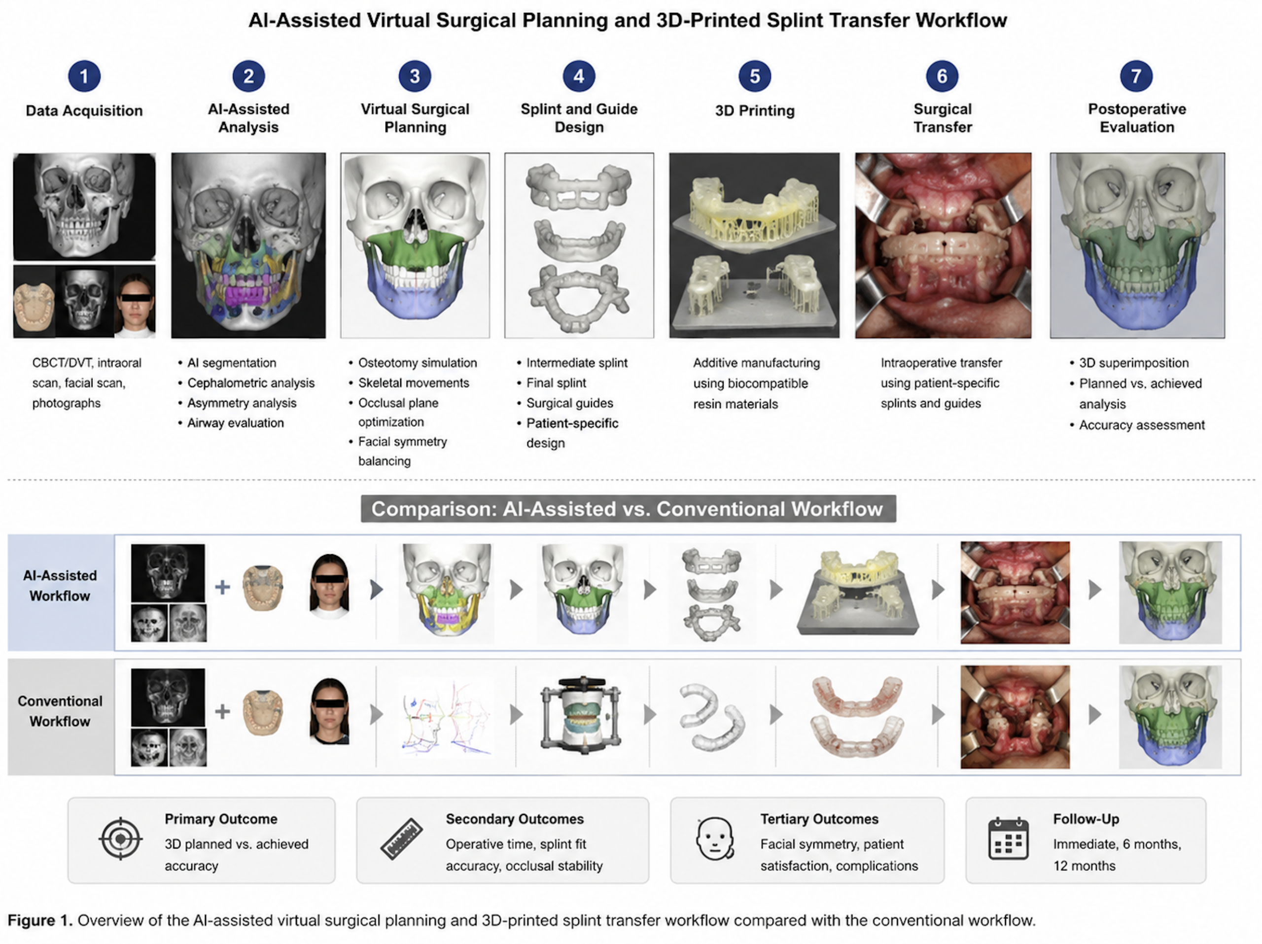
The figure illustrates the sequential integration of multimodal imaging, AI-assisted craniofacial analysis, virtual surgical planning, additive manufacturing, and intraoperative transfer using patient-specific splints and surgical guides.
3. RESULTS
A total of 124 patients completed the study protocol and were included in the final analysis. The mean patient age was 28.4 ± 6.1 years. Female patients represented 55.6% of the study population. Skeletal class III deformities represented the most common indication for surgery followed by skeletal class II deformities and facial asymmetry.
AI-assisted workflows demonstrated high clinical reliability throughout the study period. AI-assisted segmentation, cephalometric analysis, virtual planning, and additive manufacturing workflows were successfully completed in the majority of patients without major technical interruption. Minor segmentation refinements were occasionally required during early implementation phases but decreased progressively throughout the study period.
Three-dimensional superimposition analysis demonstrated significantly improved planned-versus-achieved skeletal movement accuracy in the AI-assisted cohort compared with conventional planning workflows. Maxillary repositioning demonstrated lower translational deviation values and improved angular transfer precision in the AI-assisted group. Similar improvements were observed for mandibular positioning following bilateral sagittal split osteotomy.
Rotational discrepancies including yaw, pitch, and roll deviations were significantly reduced in the AI-assisted cohort. Midline correction accuracy and postoperative facial symmetry also demonstrated improved consistency compared with conventional workflows. Three-dimensional postoperative analyses demonstrated more stable skeletal balance and lower asymmetry values following AI-assisted planning.
Operative efficiency improved in the AI-assisted group. Mean operative time was reduced due to improved intraoperative transfer precision and reduced splint adjustment requirements. Participating surgeons reported high intraoperative fit accuracy of patient-specific splints and guides.
Postoperative occlusal stability remained favorable throughout immediate postoperative evaluation and longitudinal follow-up examinations. No clinically relevant skeletal relapse was observed during 12-month follow-up analysis. Patient satisfaction and aesthetic outcome evaluation demonstrated favorable results in both groups, with a positive trend toward improved aesthetic satisfaction in the AI-assisted cohort.
Complication rates remained low throughout the study period. Minor postoperative complications included transient sensory disturbances, temporary occlusal adjustments, and isolated soft tissue irritation. No major workflow-related complication or severe digital planning failure was observed.
Comparative three-dimensional analysis revealed significantly improved planned-versus-achieved skeletal positioning accuracy in the AI-assisted cohort. Quantitative deviation analysis and representative superimposition findings are summarized in Figure 2.
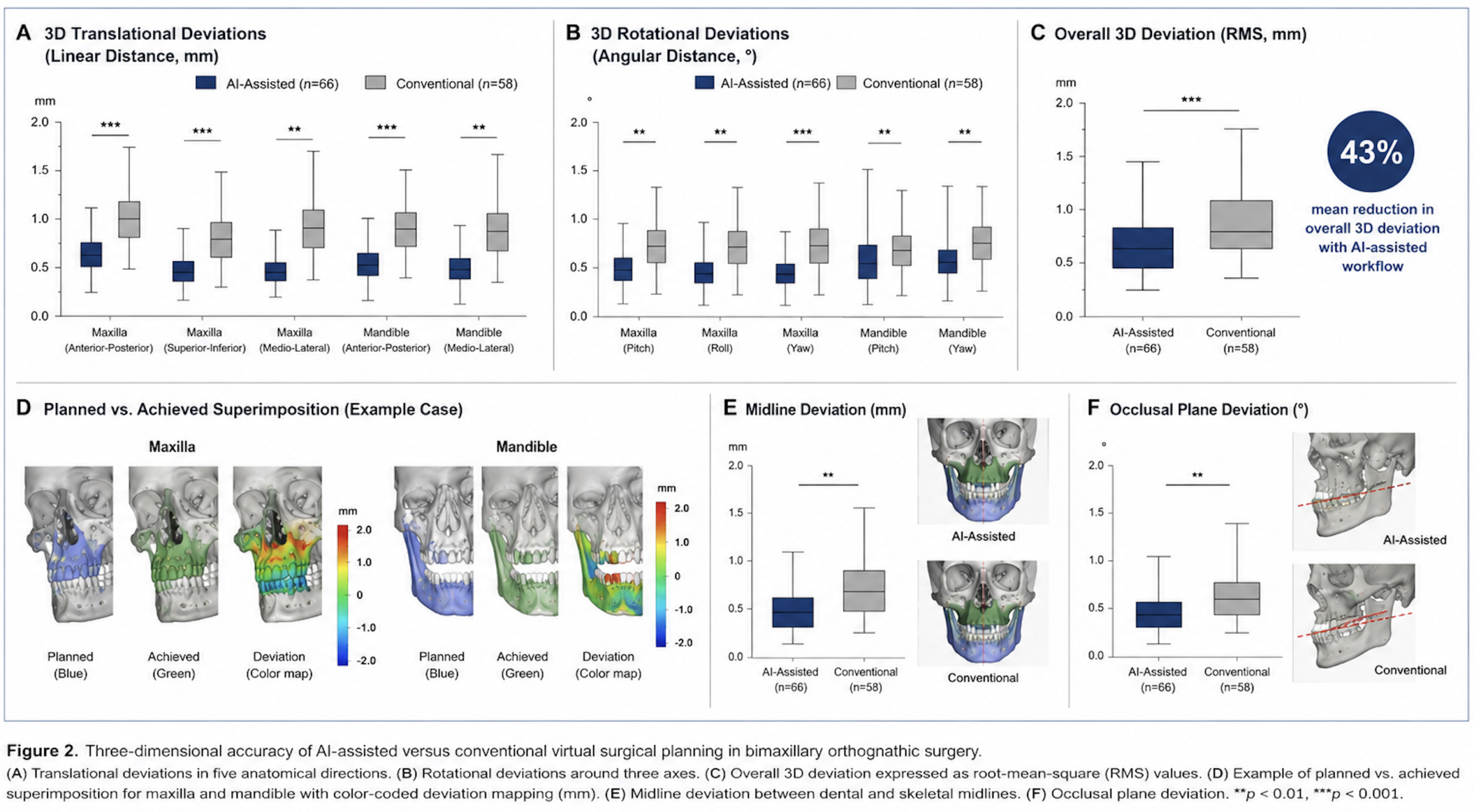
Translational and rotational deviation analyses demonstrated significantly improved skeletal transfer precision following AI-assisted planning workflows. Three-dimensional superimposition and color-coded deviation mapping revealed lower residual discrepancies together with improved midline correction and occlusal plane control compared with conventional planning methods.
4. DISCUSSION
This prospective comparative validation study demonstrates that AI-assisted virtual surgical planning combined with patient-specific 3D-printed splint transfer provides high three-dimensional surgical accuracy in bimaxillary orthognathic surgery. AI-assisted workflows demonstrated significantly reduced translational and rotational deviation values together with improved skeletal transfer precision compared with conventional planning methods.
Previous institutional investigations established the translational and clinical foundation of AI-assisted workflows in maxillofacial trauma surgery [1–13]. AI-assisted fracture detection, multicenter validation, decision support systems, surgical planning workflows, randomized controlled trials, and translational implementation studies collectively demonstrated the feasibility and clinical relevance of AI-guided personalized surgery. The present investigation extends these translational concepts into orthognathic surgery and specifically evaluates objective three-dimensional transfer accuracy.
Accurate transfer of virtual surgical planning into intraoperative skeletal positioning represents one of the most important determinants of orthognathic surgical success. Conventional workflows remain susceptible to inaccuracies related to manual cephalometric analysis, model surgery, splint fabrication, and intraoperative transfer variability. The present study demonstrated that AI-assisted segmentation, symmetry analysis, and virtual planning can significantly improve skeletal transfer precision.
Three-dimensional superimposition analysis demonstrated significantly improved surgical transfer accuracy in the AI-assisted cohort. Translational deviations across all measured directions were significantly lower for both the maxilla and mandible, while rotational discrepancies around the pitch, roll, and yaw axes were also substantially reduced. These findings resulted in a 43% reduction in overall three-dimensional deviation compared with conventional planning workflows.
The color-coded deviation maps further illustrated improved spatial congruence between planned and achieved skeletal positioning, with most residual discrepancies remaining below 1 mm in the AI-assisted group. In addition, AI-assisted planning demonstrated improved midline correction and more precise control of the occlusal plane, both of which represent critical determinants of functional stability and postoperative facial aesthetics in bimaxillary orthognathic surgery.
The reduction in translational and rotational deviation values observed in the AI-assisted cohort is clinically relevant because even small positional inaccuracies may influence postoperative occlusion, facial balance, and long-term stability. Improved yaw, pitch, and roll control may be particularly important in complex asymmetry correction and bimaxillary repositioning procedures.
The integration of additive manufacturing also contributed substantially to workflow precision. Patient-specific splints and transfer guides enabled reliable transfer of digital planning into intraoperative execution. Similar translational advantages were previously observed in AI-guided trauma surgery workflows [8–10]. The present findings confirm that personalized digital transfer concepts can also improve orthognathic surgical precision.
Postoperative symmetry outcomes and occlusal stability remained favorable throughout longitudinal follow-up. These findings suggest that AI-assisted workflows not only improve immediate surgical transfer accuracy but may also contribute to stable postoperative adaptation and skeletal balance.
The present study has several limitations. First, this was a single-center validation study performed in a specialized institution with established digital infrastructure and prior AI workflow experience. Second, long-term skeletal stability beyond 12 months was not evaluated. Third, multicenter external validation remains necessary before broader clinical generalization can be recommended.
Future investigations should evaluate randomized controlled comparisons, multicenter validation cohorts, long-term skeletal relapse analysis, automated soft tissue prediction models, and AI-assisted facial harmony optimization. The combination of artificial intelligence, virtual surgical planning, additive manufacturing, and predictive digital facial analysis may substantially influence future orthognathic surgery workflows.
5. CONCLUSION
AI-assisted virtual surgical planning combined with patient-specific 3D-printed splint transfer demonstrated high three-dimensional surgical accuracy in bimaxillary orthognathic surgery. AI-guided workflows resulted in reduced translational and rotational deviation values together with improved skeletal transfer precision and stable postoperative outcomes. These findings support the clinical reliability and translational integration of AI-guided personalized workflows in orthognathic surgery.
6. ETHICS STATEMENT
This clinical study was conducted in full accordance with the ethical principles outlined in the Declaration of Helsinki and its subsequent amendments. Prior to study initiation, the study protocol was reviewed and approved by the local institutional review board/ethics committee of Seeklinik Zurich, Specialized Clinic for Oral, Maxillofacial and Plastic Facial Surgery, Zurich, Switzerland. All participants were thoroughly informed about the purpose, procedures, potential risks, and anticipated benefits of the clinical treatment and associated digital or surgical workflows. Written informed consent was obtained from all patients prior to inclusion in the study.
Participants were informed about potential biological, surgical, technical, and procedure-related risks associated with oral and maxillofacial surgical treatment. Careful patient selection and adherence to established clinical, surgical, and digital planning protocols were implemented to minimize these risks. Patient confidentiality and data protection were rigorously maintained throughout the study, and all clinical records, imaging datasets, and digital planning files were anonymized prior to scientific analysis.
The study design ensured that no participant was exposed to undue risk, and all procedures conformed to the highest standards of contemporary clinical care. The findings of this study aim to contribute to the scientific evidence base supporting safe, effective, and translational integration of modern diagnostic, digital, and surgical workflows in oral and maxillofacial surgery.
7. CONFLICTS OF INTEREST
The authors declare no conflicts of interest related to this study.
8. FUNDING
No external funding was received for this study.
9. DATA AVAILABILITY STATEMENT
The datasets generated and analyzed during the current study are available from the corresponding author on reasonable request.
10. REFERENCES
[1] Yildirim A, Hertach R, Yildirim V. Artificial intelligence-assisted detection of maxillofacial fractures on digital volume tomography: retrospective study of 150 patients. J Med Dent. 2026;2(1):44–52.
[2] Yildirim A, Hertach R, Yildirim V. External multicenter validation of an artificial intelligence system for cone-beam CT-based detection of maxillofacial fractures: robustness across a tertiary facial trauma clinic and an independent maxillofacial practice. J Med Dent. 2026;2(1):70–81.
[3] Yildirim A, Hertach R, Yildirim V. Artificial intelligence-assisted decision support in emergency maxillofacial trauma imaging: development and validation of a CBCT-based clinical decision algorithm. J Med Dent. 2026;2(1):82–92.
[4] Yildirim A, Hertach R, Yildirim V. Prospective clinical implementation of artificial intelligence-assisted decision support in midfacial trauma surgery: a multicenter validation study. J Med Dent. 2026;2(1):93–99.
[5] Yildirim A, Hertach R, Yildirim V. Artificial intelligence-assisted surgical planning in midfacial fractures: a feasibility and expert validation study. J Med Dent. 2026;2(1):100–108.
[6] Yildirim A, Hertach R, Yildirim V. Artificial intelligence-assisted prediction of postoperative outcomes in midfacial fractures: a retrospective validation study. J Med Dent. 2026;2(1):109–117.
[7] Yildirim A, Hertach R, Yildirim V. Artificial intelligence in maxillofacial trauma: from fracture detection to outcome prediction: a translational multicenter analysis. J Med Dent. 2026;2(1):118–125.
[8] Yildirim A, Hertach R, Yildirim V. Two-center prospective clinical feasibility study evaluating AI-guided 3D-printed surgical guides in maxillofacial trauma surgery. J Med Dent. 2026;2(2):15–24.
[9] Yildirim A, Hertach R, Yildirim V. Randomized controlled trial evaluating AI-guided 3D-printed surgical guides versus conventional surgery in maxillofacial trauma. J Med Dent. 2026;2(2):25–34.
[10] Yildirim A, Hertach R, Yildirim V. Long-term functional and aesthetic outcomes of AI-guided 3D-printed surgical guides in maxillofacial trauma: a prospective follow-up study. J Med Dent. 2026;2(2):35–45.
[11] Yildirim A, Hertach R, Yildirim V. Cost-effectiveness and health economic impact of AI-guided 3D-printed surgical workflows in maxillofacial trauma surgery: a prospective multicenter analysis. J Med Dent. 2026;2(2):46–58.
[12] Yildirim A, Hertach R, Yildirim V. Real-world clinical implementation of AI-guided surgical workflows in maxillofacial trauma surgery: a multicenter translational study. J Med Dent. 2026;2(2):59–71.
[13] Yildirim A, Hertach R, Yildirim V. AI-assisted virtual surgical planning and 3D-printed splint transfer in bimaxillary orthognathic surgery. J Med Dent. 2026;2(2):72–84.
[14] Topol EJ. High-performance medicine: the convergence of human and artificial intelligence. Nat Med. 2019;25(1):44–56.
[15] Hashimoto DA, Rosman G, Rus D, Meireles OR. Artificial intelligence in surgery: promises and perils. Ann Surg. 2018;268(1):70–76.
[16] Maier-Hein L, Vedula SS, Speidel S, Navab N, Kikinis R, Park A, Eisenmann M, Feussner H, Forestier G, Giannarou S, et al. Surgical data science for next-generation interventions. Nat Biomed Eng. 2017;1:691–696.
[17] Kelly CJ, Karthikesalingam A, Suleyman M, Corrado G, King D. Key challenges for delivering clinical impact with artificial intelligence. BMC Med. 2019;17(1):195.
[18] Sendak MP, D’Arcy J, Kashyap S, Gao M, Nichols M, Corey K, Ratliff W, Balu S. A path for translation of machine learning products into healthcare delivery. NPJ Digit Med. 2020;3:16.
[19] Swennen GRJ, Mollemans W, Schutyser F. Three-dimensional treatment planning of orthognathic surgery in the era of virtual imaging. J Oral Maxillofac Surg. 2009;67(10):2080–2092.
[20] Zinser MJ, Mischkowski RA, Sailer HF, Zöller JE. Computer-assisted orthognathic surgery: feasibility study using multiple CAD/CAM surgical splints. Oral Surg Oral Med Oral Pathol Oral Radiol. 2012;113(5):673–687.
[21] Resnick CM, Dang RR, Glick SJ, Padwa BL. Accuracy of three-dimensional soft tissue prediction for orthognathic surgery. J Oral Maxillofac Surg. 2017;75(9):1971–1978.
[22] Xia JJ, Gateno J, Teichgraeber JF. New clinical protocol to evaluate craniomaxillofacial deformity and plan surgical correction. J Oral Maxillofac Surg. 2009;67(10):2093–2106.
[23] Aboul-Hosn Centenero S, Hernández-Alfaro F. 3D planning in orthognathic surgery: CAD/CAM surgical splints and prediction of soft and hard tissue results—our experience in 16 cases. J Craniomaxillofac Surg. 2012;40(2):162–168.
[24] Marchetti C, Bianchi A, Muyldermans L, Di Martino M, Lancellotti L, Sarti A. Validation of new soft tissue software in orthognathic surgery planning. Int J Oral Maxillofac Surg. 2011;40(1):26–32.
[25] Heufelder MJ, Wilde F, Pietzka S, Mascha F, Winter K, Schramm A, Rana M. Clinical accuracy of waferless maxillary positioning in computer-assisted orthognathic surgery. J Craniomaxillofac Surg. 2017;45(5):543–548.