Volume 3, Issue 2, A Prospective Three-Dimensional Clinical Study – May 05, 2026, Pages 117-126
DOI: 10.64951/jmdnt.2026.02.025
AI-Assisted Soft Tissue Prediction and Facial Symmetry Analysis Following Bimaxillary Orthognathic Surgery
Ayhan Yildirim¹*, René Hertach², Vedat Yildirim³
1 Seeklinik Zurich, Department of Oral and Maxillofacial Surgery and Clinical Research, Zurich, Switzerland
2 Center for Prosthodontics and Digital Dentistry, Zurich, Switzerland
3 Munich Center for Oral and Maxillofacial Surgery, Munich, Germany
ORCID IDs
🟢 Ayhan Yildirim: 0009-0009-2179-1802
🟢 Vedat Yildirim: 0009-0009-8664-5953
* Correspondence to: Prof. Dr. Dr. Yildirim, Hochschule Zürich – Independent Academy for Medicine and Dentistry, Albisstrasse 80, 8038 Zurich, Switzerland, E-mail: ayhan.yildirim@hs-zh.ch
Received: 12 February 2026, Revised: 25 March 2026, Accepted: 10 April 2026, Available online: 05 May 2026, Version of Record: 05 May 2026
© 2026 Journal of Medicine and Dentistry (JMDNT)
This article is published under the Creative Commons Attribution 4.0 International (CC BY 4.0) License.
You are free to share and adapt the material for any purpose, even commercially, as long as proper credit is given to the original author(s) and source.
Full license details
ABSTRACT
Background
Artificial intelligence-assisted virtual surgical planning and three-dimensional digital workflows increasingly influence modern orthognathic surgery. Although previous investigations demonstrated improved skeletal transfer accuracy and favorable long-term skeletal stability following AI-assisted orthognathic surgery, limited prospective clinical evidence exists regarding postoperative soft tissue adaptation and facial symmetry outcomes.
Objective
The aim of this prospective three-dimensional clinical study was to evaluate postoperative soft tissue prediction accuracy, facial symmetry improvement, and patient-reported aesthetic outcomes following AI-assisted virtual surgical planning in bimaxillary orthognathic surgery.
Methods
A prospective clinical study was conducted between January 2020 and December 2025 at Seeklinik Zurich, Switzerland. Patients undergoing AI-assisted bimaxillary orthognathic surgery with digital soft tissue simulation and patient-specific three-dimensional printed surgical splints were prospectively evaluated using cone-beam computed tomography, facial scanning, and standardized three-dimensional postoperative analysis. Facial symmetry, soft tissue adaptation, profile harmony, and patient-reported outcome measures were assessed during longitudinal postoperative follow-up.
Results
A total of 78 patients completed the standardized postoperative follow-up protocol. Three-dimensional postoperative analysis demonstrated high concordance between AI-predicted and clinically observed soft tissue outcomes. Significant postoperative improvement in facial symmetry indices and profile harmony was observed throughout follow-up evaluation. Patient-reported outcome measures demonstrated high postoperative satisfaction regarding facial appearance, social confidence, and aesthetic treatment outcomes. Minimal discrepancy between predicted and observed soft tissue landmarks was identified during postoperative analysis.
Conclusion
AI-assisted soft tissue prediction and three-dimensional facial symmetry analysis demonstrated favorable predictive accuracy and clinically meaningful postoperative aesthetic improvement following bimaxillary orthognathic surgery. These findings support the clinical integration of AI-assisted soft tissue simulation workflows in modern orthognathic surgery.
Keywords
artificial intelligence; orthognathic surgery; soft tissue prediction; facial symmetry; three-dimensional imaging; virtual surgical planning; digital surgery; patient-reported outcomes.
1. INTRODUCTION
The integration of artificial intelligence, three-dimensional imaging, and digital surgical workflows has substantially transformed orthognathic surgery during recent years [1–8]. Modern digital workflows increasingly combine cone-beam computed tomography, intraoral scanning, facial scanning, additive manufacturing, and artificial intelligence-assisted virtual surgical planning to improve surgical precision and postoperative predictability [9–15].
Previous investigations demonstrated favorable translational integration of artificial intelligence-assisted workflows in maxillofacial trauma surgery and orthognathic surgery [1–14]. AI-assisted fracture detection systems, multicenter validation studies, AI-guided surgical planning workflows, and randomized clinical trials collectively demonstrated improved digital workflow precision and surgical transfer accuracy in complex maxillofacial procedures [1–12].
More recently, artificial intelligence-assisted virtual surgical planning has been successfully transferred into orthognathic surgery workflows. Initial feasibility investigations demonstrated accurate three-dimensional skeletal planning and reliable additive manufacturing integration [13]. Subsequent comparative validation studies demonstrated significantly improved three-dimensional skeletal transfer accuracy following AI-assisted orthognathic surgery workflows compared with conventional planning methods [14]. Longitudinal follow-up investigations additionally demonstrated favorable postoperative skeletal stability and high patient-reported satisfaction following AI-assisted orthognathic surgery [15].
Despite these promising findings, postoperative aesthetic outcome prediction remains one of the most challenging aspects of orthognathic surgery. Although skeletal repositioning can be planned with high precision, postoperative soft tissue adaptation may vary considerably among patients because of differences in tissue thickness, muscular adaptation, age, gender, and individual facial morphology [16–22].
Accurate prediction of postoperative soft tissue adaptation and facial symmetry remains clinically important because facial aesthetics represent one of the primary motivations for orthognathic surgery [18–24]. In recent years, artificial intelligence-assisted facial analysis and three-dimensional soft tissue simulation have emerged as promising technologies for improving postoperative aesthetic prediction and patient communication [25–29].
The present prospective three-dimensional clinical study therefore aimed to evaluate postoperative soft tissue prediction accuracy, facial symmetry improvement, profile harmony, and patient-reported aesthetic outcomes following AI-assisted virtual surgical planning in bimaxillary orthognathic surgery.
2. MATERIALS AND METHODS
This prospective three-dimensional clinical study was conducted between January 2020 and December 2025 at Seeklinik Zurich, Switzerland. The study protocol followed the ethical principles of the Declaration of Helsinki and received institutional review board approval. Written informed consent was obtained from all participants prior to inclusion.
A total of 78 consecutive patients undergoing AI-assisted bimaxillary orthognathic surgery completed the prospective postoperative follow-up protocol and were included in the final analysis. Patients presenting with skeletal class II deformities, skeletal class III deformities, facial asymmetry, anterior open bite, and combined dentofacial deformities were eligible for inclusion.
Preoperative imaging included cone-beam computed tomography, intraoral digital scanning, and standardized three-dimensional facial scanning. Imaging datasets underwent artificial intelligence-assisted segmentation, cephalometric analysis, facial symmetry analysis, and virtual surgical simulation using previously validated institutional digital workflows [1–15]. Digital soft tissue simulation and postoperative facial prediction models were generated preoperatively for all patients.
Patient-specific three-dimensional printed surgical splints were fabricated using additive manufacturing technology. All patients underwent standardized bimaxillary orthognathic surgery consisting of Le Fort I osteotomy combined with bilateral sagittal split osteotomy. Additional genioplasty procedures were performed when clinically indicated.
Postoperative evaluation included immediate postoperative imaging and standardized follow-up examinations at 6 months and 12 months following surgery. Three-dimensional postoperative facial scans were superimposed onto preoperative simulation datasets to evaluate soft tissue prediction accuracy and facial symmetry outcomes.
Soft tissue landmark analysis included evaluation of nasal projection, upper lip position, lower lip projection, chin soft tissue adaptation, midline symmetry, facial width symmetry, and lower facial proportion. Translational discrepancy between predicted and observed postoperative soft tissue landmarks was measured using standardized three-dimensional analysis software.
Facial symmetry analysis evaluated bilateral soft tissue congruence and postoperative profile harmony. Patient-reported outcome measures assessed facial appearance, profile aesthetics, psychosocial confidence, social interaction, and overall postoperative satisfaction using standardized visual analog scale questionnaires.
Continuous variables were expressed as mean ± standard deviation and categorical variables as percentages. Statistical analysis was performed using parametric and non-parametric tests depending on data distribution. A p-value below 0.05 was considered statistically significant.
The complete AI-assisted digital workflow included cone-beam computed tomography acquisition, three-dimensional facial scanning, artificial intelligence-assisted virtual surgical planning, digital soft tissue simulation, additive manufacturing of patient-specific splints, and longitudinal postoperative facial symmetry analysis. The complete prospective workflow and three-dimensional postoperative evaluation protocol are illustrated in Figure 1.
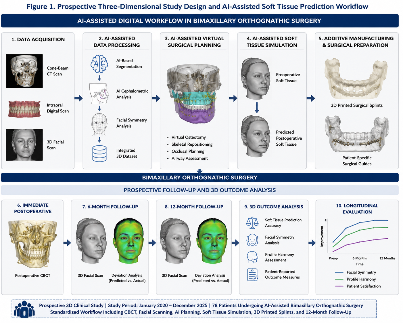
Figure 1. Prospective three-dimensional study design and AI-assisted soft tissue prediction workflow demonstrating cone-beam computed tomography integration, facial scanning, digital soft tissue simulation, additive manufacturing of patient-specific splints, and longitudinal postoperative facial symmetry analysis throughout the 12-month follow-up period.
3. RESULTS
A total of 78 patients completed the prospective follow-up protocol and were included in the final analysis. The mean patient age was 27.9 ± 6.1 years. Female patients represented 61.5% of the study population. Skeletal class III deformities represented the most common indication for surgery followed by skeletal class II deformities and facial asymmetry.
Three-dimensional postoperative analysis demonstrated high concordance between AI-predicted and clinically observed postoperative soft tissue outcomes. Mean translational discrepancy between predicted and observed soft tissue landmarks remained within clinically acceptable ranges throughout postoperative evaluation. The greatest predictive accuracy was observed in chin projection and lower facial contour adaptation.
Significant postoperative improvement in facial symmetry indices was observed following surgery. Bilateral facial congruence improved substantially during longitudinal follow-up evaluation. Midline symmetry, lower facial balance, and profile harmony demonstrated statistically significant postoperative improvement throughout the observation period.
Three-dimensional superimposition analysis demonstrated favorable correspondence between digital soft tissue simulation and clinically observed postoperative facial adaptation. Minor variation between predicted and observed postoperative nasal tip projection and upper lip adaptation was identified in isolated patients. However, overall soft tissue prediction accuracy remained highly favorable.
Patient-reported outcome measures demonstrated high levels of postoperative satisfaction. The majority of patients reported significant improvement in facial appearance, profile harmony, self-confidence, and psychosocial well-being following surgery. Overall satisfaction regarding postoperative facial aesthetics remained highly favorable throughout longitudinal follow-up evaluation.
No major workflow-related complications occurred during the study period. Minor postoperative edema and temporary sensory disturbances resolved during routine follow-up. No severe postoperative relapse or major soft tissue asymmetry requiring revision surgery was observed.
Three-dimensional postoperative facial superimposition demonstrated favorable correspondence between predicted and clinically observed postoperative soft tissue adaptation with maintained facial symmetry improvement throughout longitudinal follow-up evaluation. The complete three-dimensional soft tissue prediction analysis and postoperative facial symmetry assessment are illustrated in Figure 2.
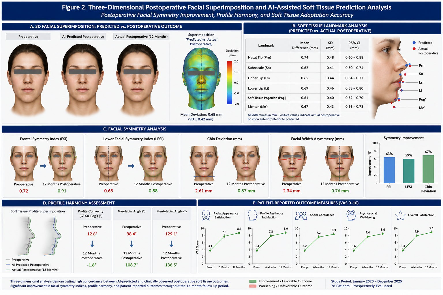
Figure 2. Three-dimensional postoperative facial superimposition and AI-assisted soft tissue prediction analysis demonstrating postoperative facial symmetry improvement, profile harmony, translational soft tissue adaptation measurements, and concordance between predicted and clinically observed postoperative facial outcomes throughout the 12-month follow-up period.
4. DISCUSSION
The present prospective three-dimensional clinical study demonstrated favorable predictive accuracy of AI-assisted soft tissue simulation and clinically meaningful postoperative facial symmetry improvement following bimaxillary orthognathic surgery.
Recent investigations increasingly demonstrated the feasibility and clinical integration of artificial intelligence-assisted workflows in oral and maxillofacial surgery [1–15]. However, most previous investigations primarily focused on skeletal transfer accuracy and digital workflow precision rather than postoperative facial aesthetics and soft tissue adaptation [13–15]. The present study therefore extends previous investigations by evaluating longitudinal postoperative facial outcomes and AI-assisted soft tissue prediction accuracy.
Postoperative facial aesthetics remain one of the most important determinants of successful orthognathic surgery [18–24]. Although accurate skeletal repositioning is essential, patient satisfaction frequently depends more strongly on postoperative facial appearance, profile harmony, and psychosocial improvement than on skeletal measurements alone [20–24].
The present findings demonstrated high concordance between predicted and observed postoperative soft tissue outcomes. Artificial intelligence-assisted soft tissue simulation demonstrated particularly favorable predictive accuracy regarding lower facial contour adaptation and chin soft tissue projection. These findings support previous investigations demonstrating the increasing reliability of modern three-dimensional facial prediction systems [25–29].
Facial symmetry improvement represented another important finding of the present investigation. Three-dimensional postoperative analysis demonstrated significant improvement in bilateral facial congruence and profile harmony throughout longitudinal follow-up evaluation. Improved facial symmetry may substantially influence postoperative psychosocial well-being and social confidence following orthognathic surgery.
Patient-reported outcome measures additionally demonstrated high postoperative satisfaction regarding facial aesthetics and psychosocial outcomes. These findings suggest that AI-assisted facial prediction may improve patient communication, preoperative expectation management, and postoperative satisfaction.
The present study has several limitations. First, this was a single-center prospective clinical study performed within a specialized digital orthognathic surgery workflow. Second, longer follow-up beyond 12 months remains necessary to evaluate long-term soft tissue adaptation and facial aging effects. Third, multicenter external validation remains necessary before broader generalization can be recommended.
Future investigations should focus on multicenter validation studies, AI-assisted automated facial harmony optimization, dynamic facial expression analysis, long-term soft tissue aging prediction, and integration of deep learning-based facial aesthetics evaluation systems.
5. CONCLUSION
AI-assisted soft tissue prediction and three-dimensional facial symmetry analysis demonstrated favorable predictive accuracy and clinically meaningful postoperative aesthetic improvement following bimaxillary orthognathic surgery. Artificial intelligence-assisted digital workflows may improve postoperative facial outcome prediction, patient communication, and aesthetic treatment planning in modern orthognathic surgery.
6. ETHICS STATEMENT
This clinical study was conducted in accordance with the ethical principles outlined in the Declaration of Helsinki and its subsequent amendments. Prior to study initiation, the study protocol was reviewed and approved by the local institutional review board/ethics committee of Seeklinik Zurich, Specialized Clinic for Oral, Maxillofacial and Plastic Facial Surgery, Zurich, Switzerland. Written informed consent was obtained from all participants prior to inclusion in the study.
7. CONFLICTS OF INTEREST
The authors declare no conflicts of interest related to this study.
8. FUNDING
No external funding was received for this study.
9. DATA AVAILABILITY STATEMENT
The datasets generated and analyzed during the current study are available from the corresponding author on reasonable request.
10. REFERENCES
[1] Yildirim A, Hertach R, Yildirim V. Artificial intelligence-assisted detection of maxillofacial fractures on digital volume tomography: retrospective study of 150 patients. J Med Dent. 2026;2(1):44–52.
[2] Yildirim A, Hertach R, Yildirim V. External multicenter validation of an artificial intelligence system for cone-beam CT-based detection of maxillofacial fractures: robustness across a tertiary facial trauma clinic and an independent maxillofacial practice. J Med Dent. 2026;2(1):70–81.
[3] Yildirim A, Hertach R, Yildirim V. Artificial intelligence-assisted decision support in emergency maxillofacial trauma imaging: development and validation of a CBCT-based clinical decision algorithm. J Med Dent. 2026;2(1):82–92.
[4] Yildirim A, Hertach R, Yildirim V. Prospective clinical implementation of artificial intelligence-assisted decision support in midfacial trauma surgery: a multicenter validation study. J Med Dent. 2026;2(1):93–99.
[5] Yildirim A, Hertach R, Yildirim V. Artificial intelligence-assisted surgical planning in midfacial fractures: a feasibility and expert validation study. J Med Dent. 2026;2(1):100–108.
[6] Yildirim A, Hertach R, Yildirim V. Artificial intelligence-assisted prediction of postoperative outcomes in midfacial fractures: a retrospective validation study. J Med Dent. 2026;2(1):109–117.
[7] Yildirim A, Hertach R, Yildirim V. Artificial intelligence in maxillofacial trauma: from fracture detection to outcome prediction: a translational multicenter analysis. J Med Dent. 2026;2(1):118–125.
[8] Yildirim A, Hertach R, Yildirim V. Artificial intelligence and additive manufacturing in maxillofacial trauma surgery: a translational framework for fracture detection, digital planning, patient-specific reconstruction, and outcome prediction. J Med Dent. 2026;2(2):1–14.
[9] Yildirim A, Hertach R, Yildirim V. AI-assisted virtual surgical planning and 3D-printed splint transfer in bimaxillary orthognathic surgery. J Med Dent. 2026;2(2):72–84.
[10] Yildirim A, Hertach R, Yildirim V. Three-dimensional accuracy of AI-assisted virtual surgical planning in bimaxillary orthognathic surgery: a prospective comparative validation study. J Med Dent. 2026;2(2):85–98.
[11] Yildirim A, Hertach R, Yildirim V. Randomized controlled trial comparing AI-assisted and conventional virtual surgical planning in bimaxillary orthognathic surgery. J Med Dent. 2026;2(2):99–110.
[12] Yildirim A, Hertach R, Yildirim V. Long-term skeletal stability and patient-reported outcomes following AI-assisted virtual surgical planning in bimaxillary orthognathic surgery. J Med Dent. 2026;2(2):111–123.
[13] Topol EJ. High-performance medicine: the convergence of human and artificial intelligence. Nat Med. 2019;25(1):44–56.
[14] Hashimoto DA, Rosman G, Rus D, Meireles OR. Artificial intelligence in surgery: promises and perils. Ann Surg. 2018;268(1):70–76.
[15] Maier-Hein L, Vedula SS, Speidel S, Navab N, Kikinis R, Park A, Eisenmann M, Feussner H, Forestier G, Giannarou S, et al. Surgical data science for next-generation interventions. Nat Biomed Eng. 2017;1:691–696.
[16] Swennen GRJ, Mollemans W, Schutyser F. Three-dimensional treatment planning of orthognathic surgery in the era of virtual imaging. J Oral Maxillofac Surg. 2009;67(10):2080–2092.
[17] Resnick CM, Dang RR, Glick SJ, Padwa BL. Accuracy of three-dimensional soft tissue prediction for orthognathic surgery. J Oral Maxillofac Surg. 2017;75(9):1971–1978.
[18] Xia JJ, Gateno J, Teichgraeber JF. New clinical protocol to evaluate craniomaxillofacial deformity and plan surgical correction. J Oral Maxillofac Surg. 2009;67(10):2093–2106.
[19] Aboul-Hosn Centenero S, Hernández-Alfaro F. 3D planning in orthognathic surgery: CAD/CAM surgical splints and prediction of soft and hard tissue results—our experience in 16 cases. J Craniomaxillofac Surg. 2012;40(2):162–168.
[20] Marchetti C, Bianchi A, Muyldermans L, Di Martino M, Lancellotti L, Sarti A. Validation of new soft tissue software in orthognathic surgery planning. Int J Oral Maxillofac Surg. 2011;40(1):26–32.
[21] Heufelder MJ, Wilde F, Pietzka S, Mascha F, Winter K, Schramm A, Rana M. Clinical accuracy of waferless maxillary positioning in computer-assisted orthognathic surgery. J Craniomaxillofac Surg. 2017;45(5):543–548.
[22] Plooij JM, Maal TJJ, Haers P, Borstlap WA, Kuijpers-Jagtman AM, Bergé SJ. Digital three-dimensional image fusion processes for planning and evaluating orthodontics and orthognathic surgery. Int J Oral Maxillofac Surg. 2011;40(4):341–352.
[23] Cevidanes LHS, Bailey LJ, Tucker GR Jr, Styner MA, Mol A, Phillips CL, Proffit WR, Turvey T. Superimposition of 3D cone-beam CT models of orthognathic surgery patients. Dentomaxillofac Radiol. 2005;34(6):369–375.
[24] Kim NK, Lee C, Kang SH, Park JW, Kim MJ, Chang YI. A three-dimensional analysis of soft and hard tissue changes after a mandibular setback surgery. Comput Methods Programs Biomed. 2006;83(3):178–187.
[25] Patcas R, Bernini DAJ, Volokitin A, Agustsson E, Rothe R, Timofte R. Applying artificial intelligence to assess the impact of orthognathic treatment on facial attractiveness and estimated age. Int J Oral Maxillofac Surg. 2019;48(1):77–83.
[26] Denadai R, Raposo-Amaral CE, Chou PY, Yen YF, Raposo-Amaral CA. Facial aesthetics and computer-assisted orthognathic surgery. Clin Plast Surg. 2019;46(4):573–593.
[27] Kaipatur NR, Flores-Mir C. Accuracy of computer programs in predicting orthognathic surgery soft tissue response. J Oral Maxillofac Surg. 2009;67(4):751–759.
[28] Hajeer MY, Ayoub AF, Millett DT, Hassan AH, Siebert JP. Current products and practices: applications of 3D imaging in orthodontics: part II. J Orthod. 2004;31(2):154–162.
[29] Schendel SA, Jacobson R, Khalessi S. 3-dimensional facial simulation in orthognathic surgery: is it accurate? J Oral Maxillofac Surg. 2013;71(8):1406–1414.