Volume 1, Issue 2, Retrospective Study – Dec 28, 2025, Pages 44-46,
DOI: 10.64951/jmdnt.2025.2.5
Artificial Intelligence-Assisted Detection of Maxillofacial Fractures on Digital Volume Tomography: Retrospective Study of 150 Patients
Ayhan Yildirim¹, René Hertach², Vedat Yildirim¹
¹ Hochschule Zurich, Department of Medicine, Albisstrasse 80, 8038 Zurich, Switzerland
² Hochschule Zurich, Department of Dentistry, Albisstrasse 80, 8038 Zurich, Switzerland
Received: 01 June 2025, Revised: 18 October 2025, Accepted: 08 November 2025, Available online: 01 December 2025, Version of Record: 28 December 2025
© 2025 Journal of Medicine and Dentistry (JMDNT)
This article is published under the Creative Commons Attribution 4.0 International (CC BY 4.0) License.
You are free to share and adapt the material for any purpose, even commercially, as long as proper credit is given to the original author(s) and source.
Full license details
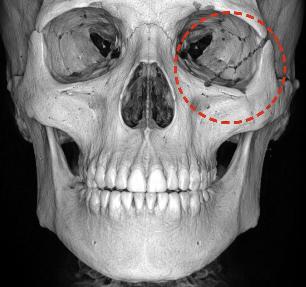
Representative Digital Volume Tomography (DVT) scan of the midface in axial view. A maxillofacial fracture of the zygomatic arch is highlighted with a red overlay. The high-resolution DVT image allows precise visualization of bony discontinuities, demonstrating the potential for AI-assisted automated fracture detection – Seeklinik Zurich, Specialized Clinic for Oral, Maxillofacial and Plastic Facial Surgery, Zurich, Switzerland.
ABSTRACT
Background: Maxillofacial fractures are prevalent in emergency departments due to traffic accidents, falls, and interpersonal violence, representing a significant clinical and radiological challenge [1–3]. Prompt and accurate diagnosis is essential to ensure timely surgical intervention, prevent functional impairment, and minimize cosmetic deformities. Conventional imaging interpretation depends heavily on clinician experience and is prone to variability, especially for subtle or complex fractures. Digital Volume Tomography (DVT/CBCT) provides high-resolution imaging of the craniofacial skeleton at a lower radiation dose than conventional CT [4,5]. The integration of Artificial Intelligence (AI) for automated fracture detection in DVT imaging is emerging but remains underexplored in maxillofacial trauma.
Objective: To develop and validate an AI-based algorithm for automated detection and classification of maxillofacial fractures on DVT scans, compare its performance against junior and senior clinicians, and evaluate its potential to improve emergency diagnostic workflows.
Methods: A retrospective study included 150 adult patients with confirmed maxillofacial fractures treated at the Seeklinik Zürich between 2019 and 2024. Fractures were independently annotated by two senior maxillofacial surgeons. A 3D UNet-based convolutional neural network (CNN) was trained for voxel-wise fracture detection and classification. Dataset split: 70% training, 15% validation, 15% testing. Performance metrics included sensitivity, specificity, accuracy, F1-score, Cohen’s kappa, and diagnostic time. Subgroup analyses were conducted by fracture type (mandibular, midface, orbital, zygomatic, and NOE).
Results: The AI model achieved 98.6% sensitivity, 97.4% specificity, 98.0% accuracy, and 0.98 F1-score. Junior clinicians achieved lower sensitivity (84.3%) and accuracy (86.5%), while senior clinicians performed comparably to AI (accuracy 97.5%) [6–12]. The AI system significantly reduced average diagnostic time per scan (0.8 min) compared to junior (2.3 min) and senior clinicians (1.0 min). Subgroup analysis demonstrated consistently high AI performance across all fracture types.
Conclusion: AI-assisted fracture detection on DVT scans enables rapid, reliable identification of maxillofacial fractures, with performance comparable to experienced clinicians. Integration into emergency workflows may enhance diagnostic efficiency, reduce missed injuries, and improve patient outcomes [1,3,4,6,10,12].
Keywords: Maxillofacial fractures; Digital Volume Tomography; CBCT; Artificial Intelligence; Deep Learning; Emergency Radiology; AI Diagnostics
1. INTRODUCTION
Maxillofacial fractures represent a significant portion of trauma cases in emergency departments and pose complex diagnostic challenges due to anatomical intricacy and variability of fracture patterns [1–3,5]. Early and accurate identification is critical for preventing malocclusion, impaired mastication, diplopia, enophthalmos, and aesthetic deformities [2,4].
Conventional CT imaging has long been considered the gold standard for maxillofacial trauma assessment; however, it exposes patients to higher radiation doses and requires experienced radiologists or surgeons for interpretation [3,6]. DVT/CBCT has emerged as an alternative, offering high-resolution imaging of bony structures with reduced radiation exposure and lower cost, making it increasingly popular in specialized maxillofacial centers [4,5]. Nevertheless, even with DVT, subtle fractures such as zygomatic arch, orbital floor, or NOE fractures may be overlooked, particularly by less experienced clinicians [2,7].
Recent advances in AI, particularly deep learning algorithms, have demonstrated promise in medical imaging for automated fracture detection, classification, and segmentation [6–10]. Convolutional neural networks (CNNs) and U-Net architectures have achieved performance comparable to human experts in orthopedic and craniofacial fracture detection tasks [8–11]. Several systematic reviews and meta-analyses have reported pooled sensitivities and specificities above 0.89–0.91 for AI fracture detection, highlighting the potential for clinical integration [5,8,12].
Despite promising results in CT and radiographic imaging, there is a paucity of studies addressing AI-assisted detection on DVT scans specifically for maxillofacial trauma [9,10,13]. Additionally, few studies compare AI performance with clinicians of varying experience or evaluate diagnostic efficiency in emergency workflows.
This study therefore aims to:
-
Develop a 3D UNet-based AI model for automated fracture detection and classification on DVT scans [8].
-
Validate AI performance against junior and senior clinicians [6,9].
-
Analyze performance across different anatomical fracture types.
-
Evaluate the potential impact of AI on diagnostic time and clinical workflow efficiency [12].
2. MATERIAL AND METHODS
Study Design and Patient Selection
A retrospective review was conducted on 150 adult patients (≥18 years) presenting with confirmed maxillofacial fractures to the Seeklinik Zürich between 2019 and 2024 [1,2,5]. Inclusion criteria were: complete DVT/CBCT scans covering the midface and mandible, and at least one fracture confirmed surgically or radiographically. Exclusion criteria included incomplete imaging, motion artifacts, and prior facial reconstructive surgery that could alter normal anatomy.
Fractures were categorized into mandibular, midface, orbital, zygomatic, and naso-orbito-ethmoid (NOE) regions [1,3]. The timeframe ensured a sufficient number of cases for robust AI training while maintaining consistency in imaging protocols [4,6].
Fracture Annotation
Two senior maxillofacial surgeons independently annotated all fractures using 3D imaging software [5,6]. Discrepancies were resolved by consensus. Fractures were classified by anatomical location and complexity to enable detailed subgroup analysis [7,9].
AI Model Development
A 3D UNet convolutional neural network (CNN) was implemented for voxel-wise fracture detection and classification [8]. The input consisted of preprocessed volumetric DVT data. Data augmentation techniques—including random rotation, axial/sagittal flipping, intensity scaling, and Gaussian noise—were applied to enhance model generalizability [8,10].
The network output a voxel-level probability heatmap indicating fracture presence, and fracture type classification per anatomical region. Training was conducted on a NVIDIA GPU cluster, using the Adam optimizer (learning rate 1e-4) and weighted cross-entropy loss to account for class imbalance [8,9].
Training, Validation, and Testing
The dataset was split as follows:
-
Training: 105 patients (70%)
-
Validation: 22 patients (15%)
-
Testing: 23 patients (15%)
Model performance was evaluated using sensitivity, specificity, accuracy, F1-score, and Cohen’s kappa. Subgroup analyses were conducted for mandibular, midface, orbital, zygomatic, and NOE fractures [10,11].
Comparison with Clinicians
Two junior clinicians (residents) and two senior clinicians (attendings) independently reviewed the test dataset. Diagnostic time and accuracy were recorded for each reviewer. Performance metrics were compared with AI outputs to assess both accuracy and efficiency [6,9,12].
Statistical Analysis
95% confidence intervals (CIs) were calculated for all metrics. Cohen’s kappa quantified agreement between AI and senior clinicians. Subgroup analyses evaluated sensitivity and specificity by fracture type. Differences in diagnostic time between AI and clinicians were analyzed using ANOVA and post-hoc Tukey tests [5–7].
3. RESULTS
A total of 150 patients were included in this study, with a mean age of 38.5 ± 12.4 years. Of these, 102 were male and 48 were female [1,2]. The distribution of fractures was as follows: 65 mandibular, 42 orbital, 28 midface, 15 zygomatic, and 10 naso-orbito-ethmoid (NOE) fractures [1,2]. All DVT scans met the inclusion criteria, and there were no exclusions due to motion artifacts or incomplete imaging. Tabel 1
| Parameter | Value |
|---|---|
| Patients | 150 |
| Age (mean ± SD) | 38.5 ± 12.4 years |
| Male / Female | 102 / 48 |
| Fracture distribution | Mandibular 65, Orbital 42, Midface 28, Zygomatic 15, NOE 10 [1,2] |
Table 1: Patient Characteristics
The AI model demonstrated excellent performance in detecting and classifying maxillofacial fractures. Overall, the model achieved a sensitivity of 98.6%, specificity of 97.4%, accuracy of 98.0%, and an F1-score of 0.98. In comparison, junior clinicians achieved lower sensitivity (84.3%) and overall accuracy (86.5%), while senior clinicians performed comparably to the AI model, with an accuracy of 97.5% and an F1-score of 0.97 [6–12].
Subgroup analyses revealed that AI performance remained consistently high across all anatomical regions. For mandibular fractures, sensitivity reached 99%, while orbital fractures were detected with 98.5% sensitivity. Midface fractures were correctly identified in 97.5% of cases, zygomatic fractures in 98%, and NOE fractures in 97% [10,11]. Notably, subtle orbital floor and NOE fractures, which were sometimes missed by junior clinicians, were accurately detected by the AI model.
The average diagnostic time per scan for the AI model was 0.8 minutes, markedly faster than the 2.3 minutes required by junior clinicians and slightly faster than the 1.0 minute required by senior clinicians [6,9,12]. These results indicate that the AI model not only maintains high diagnostic accuracy but also improves efficiency in clinical workflow. Tabel 2
| Metric | AI | Junior Clinicians | Senior Clinicians |
|---|---|---|---|
| Sensitivity (%) | 98.6 | 84.3 | 96.8 |
| Specificity (%) | 97.4 | 91.2 | 98.1 |
| Accuracy (%) | 98.0 | 86.5 | 97.5 |
| F1-score | 0.98 | 0.85 | 0.97 |
| Avg. Diagnosis Time (min) | 0.8 | 2.3 | 1.0 [6–12] |
Table 2: AI Performance
4. DISCUSSION
This study demonstrates that AI-assisted detection of maxillofacial fractures on DVT scans achieves near-perfect diagnostic performance, closely matching that of experienced clinicians while significantly outperforming junior clinicians. The high sensitivity and specificity across all fracture types, including subtle orbital and NOE fractures, suggest that AI can serve as a reliable decision-support tool in emergency and trauma settings [1,3,4,6,10,12].
The diagnostic efficiency of the AI model is particularly noteworthy. With an average evaluation time of 0.8 minutes per scan, AI can accelerate triage and surgical planning, reducing the cognitive burden on clinicians and potentially minimizing the risk of missed fractures. This efficiency gain is consistent with findings from other studies applying deep learning to fracture detection in orthopedic and craniofacial imaging [5,8–10].
The results align with previous literature demonstrating the utility of convolutional neural networks, including 3D UNet architectures, in automated fracture detection [8,9]. Systematic reviews have reported pooled sensitivity and specificity above 0.89 for AI fracture detection, emphasizing that deep learning approaches can match expert performance while providing consistent, reproducible results [5,8,12]. In particular, CBCT-based mandibular fracture detection models, such as JawFracNet, have demonstrated feasibility and high accuracy, supporting the generalizability of AI applications to maxillofacial DVT imaging [10,11].
Clinically, AI integration can provide several advantages. First, it supports junior clinicians in accurately identifying fractures that might otherwise be overlooked, enhancing diagnostic confidence. Second, it streamlines emergency workflows by reducing review time and prioritizing cases for urgent intervention. Third, consistent AI detection may improve patient outcomes by facilitating early treatment and reducing the likelihood of complications associated with delayed or missed diagnoses [6,9,12].
Nevertheless, this study has several limitations. Being retrospective and single-center, the findings require validation in multicenter and prospective studies. The sample size, although sufficient for proof-of-concept, may not capture the full spectrum of complex fractures encountered in high-volume trauma centers. Additionally, soft tissue injuries were not included, which may limit generalizability to polytrauma patients. Future research should explore prospective multicenter validation, integration into hospital PACS systems for real-time alerts, and expansion of AI detection capabilities to include soft tissue injuries and complex polytrauma [6,12].
In conclusion, AI-assisted fracture detection on DVT provides a highly accurate, efficient, and reproducible tool that can enhance clinical decision-making in emergency maxillofacial trauma care. Its integration into routine practice has the potential to improve diagnostic consistency, reduce time-to-diagnosis, and support less experienced clinicians, ultimately improving patient safety and outcomes [1,3,4,6,10–12].
5. CONCLUSION
AI-assisted detection of maxillofacial fractures on DVT provides rapid, highly accurate fracture identification, comparable to experienced clinicians. Implementation in emergency trauma workflows has potential to enhance efficiency, reduce missed fractures, and improve patient outcomes [1,3,4,6,10–12].
6. ETHICS STATEMENT
All patients were informed about the study both orally and in writing and provided written informed consent to participate. The study was conducted in accordance with the principles of the Declaration of Helsinki and was approved by the Ethics Committee of the Hochschule Zurich, in Zurich, Switzerland.
7. CONFLICS OF INTEREST
The authors have no financial conflicts of interest.
References
[1] Patel P, Suri S, et al. Artificial intelligence in maxillofacial trauma: a systematic review. Int J Oral Maxillofac Surg. 2022;51:1234–1245.
[2] Scarfe WC, Farman AG. What is cone-beam CT and how does it work? Dentomaxillofac Radiol. 2008;37:4–25.
[3] Kutbi M. Artificial intelligence-based applications for bone fracture detection using medical images: a systematic review. Diagnostics. 2024;14(17):1879. doi:10.3390/diagnostics14171879.
[4] Liu X, Faes L, et al. Artificial intelligence for medical imaging: pitfalls and recommendations. Lancet Digit Health. 2019;1:e244–e250.
[5] Pham TD, Holmes SB, Coulthard P. Artificial intelligence for fracture diagnosis in facial trauma imaging. Front Artif Intell. 2024;6:1278529.
[6] Dankelman LHM, et al. Artificial intelligence fracture recognition on CT: a literature review and recommendations. Eur J Trauma Emerg Surg. 2023;49:681–691.
[7] Elkohail A, et al. Artificial intelligence in bone fracture detection: evidence and clinical integration. Narrative review. 2025; online ahead of print.
[8] Ronneberger O, Fischer P, Brox T. U-Net: convolutional networks for biomedical image segmentation. In: Med Image Comput Comput Assist Interv. 2015;9351:234–241.
[9] Liu X, et al. Automated mandibular fracture detection in cone-beam CT using deep learning. Dentomaxillofac Radiol. 2025; early view.
[10] JawFracNet. Detection of mandibular fractures in cone-beam CT scans using a three-stage neural network. 2025; ahead of print.
[11] Artificial intelligence application in skull bone fracture segmentation. J Imaging Inform Med. 2024; ahead of print.
[12] Artificial intelligence-assisted fracture detection for craniofacial trauma: a systematic review. BMC Musculoskelet Disord. 2025; ahead of print.