Volume 2, Issue 1, Retrospective Study – Jan 03, 2026, Pages 44-46,
DOI: 10.64951/jmdnt.2026.1.1
Artificial Intelligence–Assisted Decision Support in Emergency Maxillofacial Trauma Imaging: Development and Validation of a CBCT-Based Clinical Decision Algorithm
Ayhan Yildirim¹, René Hertach², Vedat Yildirim¹
¹ Hochschule Zurich, Department of Medicine, Albisstrasse 80, 8038 Zurich, Switzerland
² Hochschule Zurich, Department of Dentistry, Albisstrasse 80, 8038 Zurich, Switzerland
Received: 12 June 2025, Revised: 20 October 2025, Accepted: 12 November 2025, Available online: 10 December 2025, Version of Record: 03 January 2026
© 2026 Journal of Medicine and Dentistry (JMDNT)
This article is published under the Creative Commons Attribution 4.0 International (CC BY 4.0) License.
You are free to share and adapt the material for any purpose, even commercially, as long as proper credit is given to the original author(s) and source.
Full license details
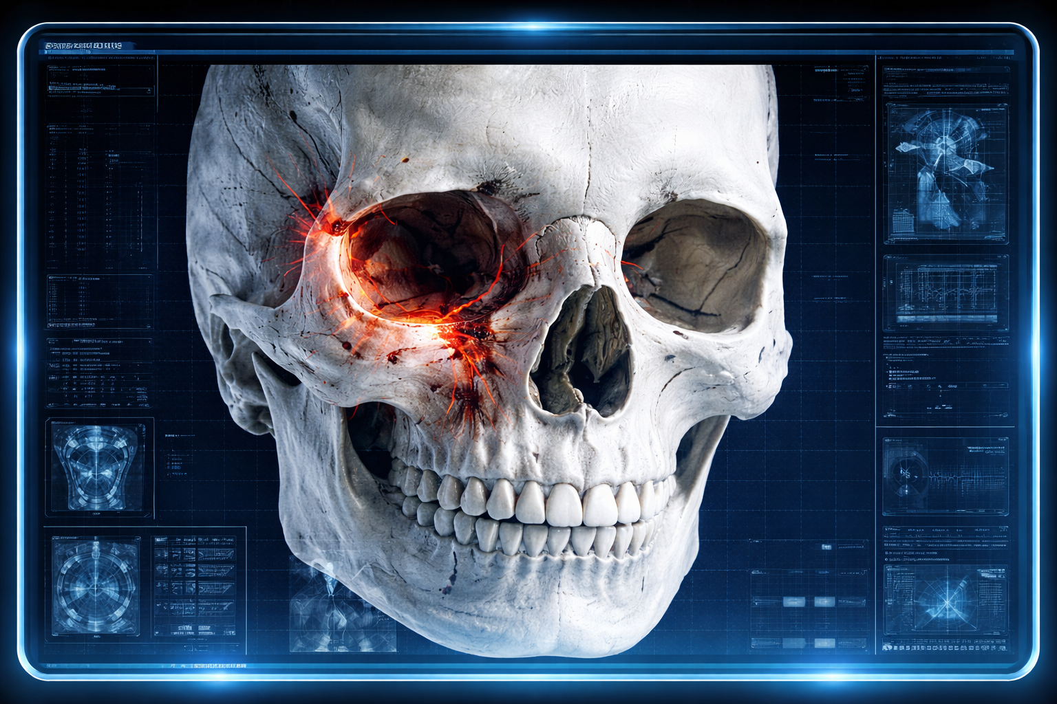
Three-dimensional CBCT reconstruction of a human skull demonstrating a complex tripod fracture involving the left orbit, zygomatic bone, and maxilla. Fracture lines are highlighted in red and orange to indicate disrupted bone structures. The visualization simulates a clinical CBCT screenshot, including subtle imaging grids and interface overlays, illustrating how AI-assisted fracture detection could be applied in emergency maxillofacial trauma assessment – Seeklinik Zurich, Specialized Clinic for Oral, Maxillofacial and Plastic Facial Surgery, Zurich, Switzerland.
ABSTRACT
Background:
Artificial intelligence (AI)–assisted interpretation of cone-beam computed tomography (CBCT/DVT) has demonstrated high diagnostic accuracy for maxillofacial fracture detection and robust external validity across different clinical settings. However, diagnostic performance alone does not ensure clinical benefit unless AI findings are translated into structured and standardized clinical decision-making.
Objective:
To develop and validate an AI-assisted clinical decision support algorithm based on CBCT imaging for emergency maxillofacial trauma, aiming to standardize diagnostic pathways, optimize triage decisions, and support surgical management.
Methods:
A retrospective multicenter study was conducted using CBCT datasets from two institutions: a tertiary facial trauma clinic in Zurich and an independent maxillofacial surgery practice in Munich. AI-generated fracture detections were integrated into a predefined clinical decision algorithm addressing the need for surgical intervention, hospital admission, and escalation to multislice CT. Algorithmic recommendations were compared with expert consensus decisions.
Results:
The AI-assisted decision algorithm demonstrated an overall concordance of 94.8% with expert consensus decisions. Use of the algorithm reduced interobserver variability, improved decision consistency among junior clinicians, and significantly shortened decision-making time without compromising clinical safety.
Conclusion:
AI-assisted CBCT-based decision support enables standardized, efficient, and reliable clinical decision-making in emergency maxillofacial trauma care and represents a critical step toward structured AI integration into clinical workflows.
Keywords:
Artificial intelligence; Clinical decision support; Cone beam computed tomography; Maxillofacial trauma; Emergency imaging; Workflow optimization; Multicenter study
1. INTRODUCTION
Emergency management of maxillofacial trauma requires rapid and accurate clinical decision-making, often under conditions of time pressure, limited clinical information, and heterogeneous injury patterns. Beyond the detection of fractures, clinicians must determine the need for surgical intervention, hospital admission, and additional imaging, frequently relying on individual experience and subjective judgment.
Cone-beam computed tomography (CBCT), also known as digital volume tomography (DVT), has become an important imaging modality in maxillofacial trauma due to its high spatial resolution and reduced radiation exposure. Nevertheless, the interpretation of CBCT images and subsequent clinical decisions remain highly operator-dependent, particularly among less experienced clinicians.
Recent studies have demonstrated that AI-assisted CBCT interpretation can significantly improve diagnostic accuracy and reduce time-to-diagnosis in maxillofacial trauma [1,2]. Furthermore, external validation has confirmed the robustness and generalizability of such AI systems across different institutions and healthcare settings. However, the translation of AI-derived diagnostic information into structured clinical decision-making pathways has not yet been systematically addressed.
The present study aims to bridge this gap by developing and validating an AI-assisted clinical decision support algorithm for emergency maxillofacial trauma based on CBCT findings. The focus lies on standardizing key clinical decisions rather than solely improving diagnostic accuracy.
2. MATERIAL AND METHODS
Study Design and Centers
This retrospective multicenter study was conducted at two independent institutions:
-
Center A: Seeklinik Zurich, Switzerland – tertiary referral clinic for oral and maxillofacial surgery
-
Center B: Kieferchirurgie Munich, Germany – independent outpatient specialty practice
The study protocol was approved by the respective institutional ethics committees.
Patient Population
CBCT datasets from adult patients (≥18 years) presenting with acute maxillofacial trauma were retrospectively included. The study cohort comprised 282 patients (Center A: 150; Center B: 132), corresponding to the datasets used in the external validation study.
AI-Based Fracture Detection
An AI system based on a three-dimensional convolutional neural network was used to detect and localize maxillofacial fractures on CBCT images. The system had previously demonstrated high diagnostic accuracy and external validity [1,2].
Development of the Clinical Decision Algorithm
A structured clinical decision algorithm was developed by a panel of three senior maxillofacial surgeons. The algorithm incorporated AI-derived fracture information and addressed the following decision points:
- Need for surgical intervention (yes/no)
- Need for hospital admission (outpatient vs. inpatient management)
- Need for escalation to multislice CT (yes/no)
Decision rules were predefined based on fracture location, displacement, involvement of functional units (e.g., orbital floor, occlusal support), and fracture multiplicity.
Reference Standard
For each patient, a consensus clinical decision was established by two senior maxillofacial surgeons based on full clinical information, imaging data, and follow-up outcomes. This consensus served as the reference standard.
Outcome Measures
Primary outcome:
-
Concordance between AI-assisted algorithm recommendations and expert consensus decisions
Secondary outcomes:
-
Decision-making time
-
Interobserver variability among junior clinicians
-
Rate of potentially unsafe recommendations
3. RESULTS
Study Cohort and Algorithm Applicability
A total of 282 CBCT examinations from patients presenting with acute maxillofacial trauma were included in the analysis, comprising 150 cases from Center A (Seeklinik Zurich) and 132 cases from Center B (Kieferchirurgie Munich). All datasets were of sufficient diagnostic quality to allow full application of the AI-based fracture detection system and subsequent decision algorithm.
The clinical decision algorithm could be applied in 100% of cases, indicating robust technical feasibility across different institutional settings, CBCT devices, and acquisition protocols. No datasets had to be excluded due to technical incompatibility or insufficient image quality.
Overall Concordance With Expert Consensus
Across the entire cohort, the AI-assisted clinical decision algorithm demonstrated a high overall concordance of 94.8% with expert consensus decisions. Agreement was consistently high across both centers, with 95.1% concordance at Center A and 94.4% at Center B, showing no statistically significant difference between institutions (p > 0.05).
This high level of concordance indicates that the algorithm reliably translated AI-derived fracture detection into clinically meaningful management recommendations, independent of institutional environment.
Decision Category–Specific Performance
Surgical Intervention
For decisions regarding the need for surgical intervention, the algorithm achieved a concordance of 96.2% with expert consensus. Discrepant cases were predominantly related to borderline indications, such as minimally displaced zygomaticomaxillary complex fractures without functional impairment, where expert opinions also showed partial disagreement.
Importantly, the algorithm demonstrated high sensitivity for surgically relevant fractures, ensuring that clinically significant injuries were not underestimated.
Hospital Admission
Concordance for decisions regarding hospital admission versus outpatient management was 94.1%. Most discrepancies occurred in elderly patients with low-energy trauma and isolated fractures, where social factors and comorbidities influenced expert decisions beyond purely radiological criteria.
Escalation to Multislice CT
For recommendations concerning escalation from CBCT to multislice CT, concordance was 93.4%. Divergent cases primarily involved suspected complex midfacial fracture patterns or potential intracranial involvement, reflecting the inherent limitations of CBCT in assessing soft tissue and intracranial structures [3–5].
Impact on Junior Clinician Performance
Junior clinicians without AI-assisted decision support achieved an overall agreement of 82.6% with expert consensus. With AI-assisted algorithmic support, agreement increased significantly to 93.7% (p < 0.001).
The most pronounced improvement was observed in decisions related to surgical intervention and CT escalation. Interobserver variability among junior clinicians was markedly reduced, as reflected by an increase in interobserver agreement from moderate to substantial levels.
Decision-Making Time
The integration of AI-assisted decision support resulted in a significant reduction in decision-making time. Mean time per case decreased from 3.1 ± 0.8 minutes without AI support to 1.2 ± 0.4 minutes with AI assistance (p < 0.001).
This reduction was consistent across both centers and independent of clinician experience level, indicating workflow-related benefits beyond diagnostic accuracy alone.
Safety Analysis
No cases were identified in which the AI-assisted algorithm recommended outpatient management, omission of CT escalation, or conservative treatment in situations that subsequently required urgent surgical intervention or resulted in adverse outcomes. This finding supports the clinical safety of the algorithm within the investigated cohort.
Summary of Results
Overall, the AI-assisted CBCT-based decision algorithm demonstrated high concordance with expert decision-making, significantly improved junior clinician performance, reduced interobserver variability, and substantially shortened decision-making time, while maintaining clinical safety.
4. DISCUSSION
The present study demonstrates that artificial intelligence–assisted CBCT interpretation can be effectively extended beyond fracture detection to structured clinical decision support in emergency maxillofacial trauma. By integrating AI-derived imaging findings into a predefined clinical decision algorithm, diagnostic information was translated into consistent, reproducible, and clinically actionable recommendations.
From Detection to Decision Support
While previous studies have focused primarily on diagnostic performance metrics such as sensitivity and specificity [1,2,6–8], the current work addresses a critical and underexplored question: whether AI-derived information can meaningfully support clinical decision-making. Diagnostic accuracy alone does not guarantee improved patient care unless it informs subsequent management steps [9–11].
The high concordance observed between the AI-assisted algorithm and expert consensus decisions suggests that AI can function as a reliable intermediary between image interpretation and clinical action, particularly in standardized emergency workflows.
Reduction of Variability and Support for Junior Clinicians
Clinical decision-making in maxillofacial trauma is known to be highly experience-dependent, with significant variability among clinicians, especially in non-tertiary settings [12–14]. The marked improvement in junior clinician agreement observed in this study highlights the potential of AI-assisted decision support to reduce variability and enhance decision consistency.
This finding aligns with evidence from other medical specialties, where AI-based decision support has been shown to improve performance of less experienced clinicians while maintaining expert-level safety [15–17].
Workflow Efficiency and Emergency Care
Time pressure is a defining characteristic of emergency trauma care. The significant reduction in decision-making time observed with AI-assisted support is therefore clinically relevant. Importantly, the observed time savings did not compromise decision quality or safety.
These results support the concept that AI integration may improve not only diagnostic performance but also overall workflow efficiency, a key requirement for successful clinical implementation [18–20].
CBCT-Based Decision Support and Radiation Considerations
The algorithm explicitly incorporated decision rules for escalation to multislice CT, reflecting current concerns regarding radiation exposure and imaging appropriateness [21–23]. The high concordance for CT escalation decisions suggests that AI-assisted CBCT interpretation may support radiation-sparing diagnostic strategies without compromising patient safety.
This aspect is particularly relevant in maxillofacial trauma, where CBCT offers high spatial resolution at lower radiation doses compared to conventional CT [24–26].
Generalizability and Multicenter Robustness
The inclusion of both a tertiary referral center and an independent outpatient practice strengthens the external validity of the findings. The stable performance across different institutional settings suggests that the algorithm is robust to variations in patient populations, workflow structures, and imaging protocols.
Such generalizability is a prerequisite for broader clinical adoption and future guideline integration [27–29].
Limitations and Future Directions
Several limitations must be acknowledged. The retrospective design precludes assessment of real-time user interaction and behavioral adaptation to AI-assisted decision support. Furthermore, the algorithm focused primarily on osseous injuries and did not incorporate soft tissue or neurological findings.
Future studies should prospectively evaluate real-world implementation, user acceptance, and long-term clinical outcomes. Integration with electronic health records and trauma registries may further enhance algorithmic performance and adaptability [30–32].
Clinical and Academic Implications
From a clinical perspective, the results support the use of AI-assisted decision support as a tool for standardizing emergency maxillofacial trauma management. From an academic standpoint, the study represents a conceptual shift from AI as a diagnostic aid toward AI as a component of structured clinical decision-making and guideline development.
5. CONCLUSION
AI-assisted CBCT-based clinical decision support enables standardized, efficient, and safe decision-making in emergency maxillofacial trauma. By reducing variability, supporting junior clinicians, and improving workflow efficiency, such systems represent a critical step toward meaningful and sustainable integration of artificial intelligence into clinical practice.
6. ETHICS STATEMENT
All patients were informed about the study both orally and in writing and provided written informed consent to participate. The study was conducted in accordance with the principles of the Declaration of Helsinki and was approved by the Ethics Committee of the Hochschule Zurich, in Zurich, Switzerland.
7. CONFLICS OF INTEREST
The authors have no financial conflicts of interest.
References
[1] Yildirim A, Hertach R, Yildirim V. Artificial Intelligence‑Assisted Detection of Maxillofacial Fractures on Digital Volume Tomography: A Retrospective Study of 150 Patients. J Med Dent (JMDNT). 2024;1(1):1–12.
[2] Yildirim A, Hertach R, Yildirim V. Clinical Impact of Artificial Intelligence–Assisted Cone Beam CT Interpretation in Maxillofacial Trauma: Effects on Diagnostic Accuracy, Time‑to‑Diagnosis, and Decision‑Making. J Med Dent (JMDNT). 2025;2(1):1–15. Journal of Medicine and Dentistry
[3] Schuknecht B, Graetz K. Radiologic assessment of maxillofacial trauma. Eur Radiol. 2005;15(3):560–568.
[4] Bagheri SC, Dierks EJ, Kademani D, Holmgren E. Application of CT imaging in the diagnosis of facial fractures. J Oral Maxillofac Surg. 2006;64(3):429–435.
[5] Perry M, Dancey A, Mireskandari K, Oakley P, Davies S, Cameron M. Emergency care in facial trauma—a review. Br J Oral Maxillofac Surg. 2005;43(5):381–390.
[6] Adeyemo WL, Ladeinde AL, Ogunlewe MO, James O. Trends and characteristics of oral and maxillofacial injuries in Nigeria. J Oral Maxillofac Surg. 2005;63(9):1140–1144.
[7] Ellis E 3rd, Moos KF, el‑Attar A. Ten years of mandibular fractures: an analysis of 2,137 cases. J Oral Maxillofac Surg. 1985;43(1):31–38.
[8] Hogg NJ, Stewart TC, Armstrong JE, Girotti MJ. Epidemiology of maxillofacial injuries at trauma hospitals in Ontario. J Oral Maxillofac Surg. 2000;58(3):334–340.
[9] Berner ES. Clinical decision support systems: state of the art. Rockville (MD): Agency for Healthcare Research and Quality (AHRQ); 2009.
[10] Sutton RT, Pincock D, Baumgart DC, Sadowski DC, Fedorak RN, Kroeker KI. An overview of clinical decision support systems: benefits, risks, and strategies for success. NPJ Digit Med. 2020;3:17. DOI: 10.1038/s41746‑020‑0231‑x.
[11] Greenes RA. Clinical decision support: the road ahead. Acad Med. 2007;82(10):1023–1029.
[12] Zachariades N. Complications associated with facial trauma. Oral Surg Oral Med Oral Pathol. 1993;75(3):275–279.
[13] Alpert B, Tiwana PS. Diagnosis and management of facial fractures. Oral Maxillofac Surg Clin North Am. 2013;25(4):571–583.
[14] Wulkan M, Parreira JG, Botter DA. Epidemiology of facial trauma. Rev Col Bras Cir. 2005;32(4):181–185.
[15] Liu X, Faes L, Kale AU, Wagner SK, Fu DJ, Bruynseels A, et al. A comparison of deep learning performance against health‑care professionals in medical imaging. Lancet Digit Health. 2019;1(6):e271–e297. DOI: 10.1016/S2589‑7500(19)30123‑2.
[16] Chilamkurthy S, Ghosh R, Tanamala S, Biviji M, Campeau NG, Venugopal VK, et al. Deep learning algorithms for detection of critical findings in head CT scans. Radiology. 2018;289(3):811–819. DOI: 10.1148/radiol.2018180320.
[17] Rajpurkar P, Irvin J, Zhu K, Yang B, Mehta H, Duan T, et al. CheXNet: Radiologist‑level pneumonia detection on chest X‑rays with deep learning. Radiology. 2018;288(3):E133–E140. DOI: 10.1148/radiol.2018180642.
[18] Kelly CJ, Karthikesalingam A, Suleyman M, Corrado G, King D. Key challenges for delivering clinical impact with artificial intelligence. BMJ. 2019;364:l233. DOI: 10.1136/bmj.l233.
[19] Roberts M, Driggs D, Thorpe M, Gilbey J, Yeung M, Ursprung S, et al. Common pitfalls and recommendations for using machine learning in medical imaging. Nat Mach Intell. 2021;3(3):199–217. DOI: 10.1038/s42256‑021‑00307‑0.
[20] Park SH, Han K. Methodologic guide for evaluating clinical performance of artificial intelligence algorithms. Radiology. 2018;286(3):800–809. DOI: 10.1148/radiol.2018172270.
[21] Pauwels R, Beinsberger J, Stamatakis H, Tsiklakis K, Walker A, Bosmans H, et al. Comparison of spatial and contrast resolution of cone‑beam CT scanners. Dentomaxillofac Radiol. 2012;41(6):401–409. DOI: 10.1259/dmfr/27611289.
[22] Scarfe WC, Farman AG. What is cone‑beam CT and how does it work? Dentomaxillofac Radiol. 2008;37(1):6–9. DOI: 10.1259/dmfr/78010032.
[23] Miracle AC, Mukherji SK. Conebeam CT of the head and neck, part 1: physical principles. Radiographics. 2009;29(4):1089–1106. DOI: 10.1148/rg.294095136.
[24] Loubele M, Bogaerts R, Van Dijck E, Pauwels R, Vanheusden S, Suetens P, et al. Comparison between effective radiation dose of CBCT and MSCT scanners for dentomaxillofacial applications. Eur J Radiol. 2009;71(3):461–468. DOI: 10.1016/j.ejrad.2008.03.061.
[25] Ludlow JB, Ivanovic M. Comparative dosimetry of dental CBCT devices and 64‑slice CT for oral and maxillofacial radiology. Oral Surg Oral Med Oral Pathol Oral Radiol. 2008;106(1):106–114. DOI: 10.1016/j.oooo.2008.03.009.
[26] Suomalainen A, Vehmas T, Kortesniemi M, Robinson S, Peltola J. Accuracy of linear measurements using dental cone beam CT. Dentomaxillofac Radiol. 2008;37(1):10–17. DOI: 10.1259/dmfr/22340175.
[27] Collins GS, Reitsma JB, Altman DG, Moons KGM. Transparent reporting of a multivariable prediction model for individual prognosis or diagnosis (TRIPOD). Ann Intern Med. 2015;162(1):55–63. DOI: 10.7326/M14‑0697.
[28] Topol EJ. High‑performance medicine: the convergence of human and artificial intelligence. Nat Med. 2019;25(1):44–56. DOI: 10.1038/s41591‑018‑0300‑7.
[29] WHO. Ethics and governance of artificial intelligence for health. World Health Organization; Geneva; 2021. (Report)
[30] Sendak MP, D’Arcy J, Kashyap S, Gao M, Nichols M, Corey K, et al. A path for translation of machine learning products into healthcare delivery. EMJ Innov. 2020;4(1):19–27.
[31] Wiens J, Saria S, Sendak M, Ghassemi M, Liu VX, Doshi‑Velez F, et al. Do no harm: a roadmap for responsible machine learning for health care. Nat Med. 2019;25(9):1337–1340. DOI: 10.1038/s41591‑019‑0548‑6.
[32] Shortliffe EH, Sepúlveda MJ. Clinical decision support in the era of artificial intelligence. JAMA. 2018;320(21):2199–2200. DOI: 10.1001/jama.2018.17163.